
Family Card - Person Sheet
Family Card - Person Sheet
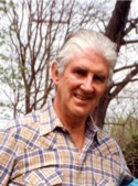
NameJohn Michael BEDFORD 
 , 69
, 69
, 69Death24 February 2018, 1901 Walnut Street (12F), Philadelphia, PA 19103, USA65 Age: 85
FatherWilliam Ritchie BEDFORD Bill , 195 (1903-1978)
MotherIda Maria MORRIS , 68 (1910-2010)
Notes for John Michael BEDFORD
Email from Kate Duckering to James Rutherford about Mike:
Hi James
Just off out for the day but I can tell you a bit of what I know and I can try and check other stuff later tonight by calling Anne Lord to see if she knows/remembers anything.
Parents were William Ritchie Bedford (known as Bill Bedford) and Ida Maria Morris - they were married on 8th August 1931 and John Michael Bedford was clearly conceived very soon after as he was duly born 9 months later on 21st May 1932 in Sheffield, Yorkshire where they were living. Mike (as he was always known in the family) was always very proud of his Yorkshire roots which made him eligible to play cricket for Yorkshire if he so wanted.
My mother Susan (Sue) Bedford was born on 14th March 1934, also in Sheffield and then the family of four was complete.
My grandparents (Bill and Ida) were not wealthy people but worked hard to ensure that both children had a good education. Bill was a draughtsman working in companies that made machinery, and Ida was a housewife - doing some voluntary work during the war. The family moved a fair amount around the country so they never owned their own property but rented. The war years covered 1939 - 1945 and during that time they found themselves living in Ruislip which is on the western outskirts of London - so very near the bombing. Ida and the children were evacuated first to stay with the Driver cousins in Yorkshire, then to a farm in Cumbria (English Lake District) and to Whitley Bay in Northumberland - don’t know the dates. Then they went home to Ruislip as they missed being together and meanwhile Bill had got a simple air raid shelter (‘Anderson’ shelter) fixed up in their home.
Mike was a very active child - my grandmother Ida said he was ‘always busy’ - and as they moved around he was very good at adopting the local accent - so as to ‘blend in’. We have evidence that they were living in Bristol in 1944 and Mike was attending Bristol Grammar School. He went to Blundells which is a small public (you would say private) school in Tiverton, Devon from 1945 to complete his high school education. Mike thrived at this school and did very well academically and in the school sports - athletics, cricket and rugby.
My grandparents decided that Mike would enjoy being a veterinary surgeon, they imagined he would like the country lifestyle and I guess he must have liked animals and this is the path he took when he studied veterinary science at Cambridge University after he did his National Service which for some time after the war years was compulsory. After a while working in this field he found a different career pathway working in reproductive research which took him to the States where he settled. At one stage he was talking to London Zoo about the possibility of a job there - I believe the head of the Zoo - but he decided to stay in the States and continue in medical research.
Love Kate
CURRICULUM VITAE: JOHN MICHAEL BEDFORD, Vet. M.B., Ph.D.
BIRTH DATE: May 21, 1932
BIRTH PLACE: Sheffield, England.
CITIZENSHIP: British/U.S. naturalized: 1993.
STATUS: Married : Rita Reinhardt-Bedford
ADDRESS:
HOME: 1901 Walnut St (12F), Rittenhouse Plaza, Philadelphia, PA, 19103, U.S.A.
WORK: Weill College of Medicine of Cornell University, 1300 York Avenue, (Box 30), New York, NY 10021
Tel/ 267 639 3412; E mail: mbedford@med.cornell.edu
MILITARY SERVICE
1950-1952 Lieutenant, Royal Corps of Signals
EDUCATION
1952-1958 Cambridge University, Sidney Sussex College
1955, B.A., 1958, M.A. Natural Sciences (Physiology, Zoology, Biochemistry, Pathology)
66POSTGRADUATE TRAINING
1958 Vet. M.B., Veterinary Medicine and Surgery, Cambridge University,
1961-1965 Ph.D., Physiology, University of London Faculty of Science, (Supervisor: Prof. E.C. Amoroso)
ACADEMIC APPOINTMENTS
1958-1959 Fellow, Department of Veterinary Surgery, Bristol University, (Lectured in Surgical Pathology, Surgical Technique)
1959-1961 Scientist (with Dr. M.C. Chang), The Worcester Foundation for Experimental Biology, Shrewsbury, Massachusett
1961-1966 Lecturer, Department of Physiology, The Royal Veterinary College, University of London
1965 Established Teacher in Physiology, University of London)
1966-1967 Scientist, The Worcester Foundation for Experimental Biology
1967-1970 Assistant Professor of Anatomy, Department of Anatomy, Columbia University College of Physicians and Surgeons
1970-1972 Associate Professor of Anatomy, Department of Anatomy, Columbia University College of Physicians and Surgeons
1972-2000 Professor of Reproductive Biology, Department of Obstetrics and Gynecology, Cornell University Medical College
1972-2000 Professor of Cell Biology and Anatomy, Department of Anatomy, Cornell University Medical College
1981-2000 Percy and Harold Uris Professor of Reproductive Biology, Cornell University Medical College
2000 - Professor Emeritus of Reproductive Biology in Obstetrics and Gynecology
1986-1990 Director, In Vitro Fertilization Laboratories, Cornell University Medical College
FORMAL TEACHING. 1958: Lecture courses on surgical pathology, and on surgical technique, Bristol Univ. Vet School. 1961-1966. Lectures in Physiology, Royal Vet. College. 1967-1972: Lectures to 1st year medical students in Gross Anatomy and Cell Biology (Columbia College of Physicians and Surgeons); 1972–1979: lectures to 1st year medical class in Gross Anatomy and Cell Biology; 1975–1979 special course in biology of conception for 1st year students; 1980–2000: lectures to 1st year medical students on mechanisms of conception and generation of the embryo (Physiology/Endocrinology - Cornell Medical College). 1996 lectures to medical students at Cornell/Qatar
HONORS/AWARDS
1954 Elected to the Hawks Athletic Club: Cambridge University
1959 Fitzwygram Prize : Royal College of Veterinary Surgeons
1982 Serono Award for Distinguished Contributions to Andrology, American Association of Andrology
1992 Distinguished Visiting Scholar : The University of Adelaide, South Australia
1995 Foreign Special Visiting Professor, Kyushu University, Fukuoka, Japan.
1995 Bruce Stewart Memorial Lecturer: American Society of Reproductive Medicine
Distinguished Andrologist Award : American Society of Andrology
1998 Keynote Speaker – Joint Meeting of German, Swiss and Austrian Reproduction Societies
1999 The Marshall Medal: British Society for the Study of Fertility.
2001 Keynote Speaker – Alpha Society of Human Embryologists, New York Meeting
EDITORIAL ACTIVITIES
1971-1975 Editorial Board, Journal of Experimental Zoology
1974-1978 Editorial Board, Biology of Reproduction
1978-1985 Editorial Board, American Journal of Anatomy
1982-1985 Editorial Board, Journal of Andrology
1983-1988 Editorial Board, Journal of Assisted Reproduction and Genetics
1992-1999 Editorial Board, Zygote
1998-2007 Editorial Board, Journal of Reproduction and Fertility (Reproduction)
ADVISORY
1970-1974 Study Section for Reproductive Biology (grant application evaluation), National Institutes of Health, Washington, D.C.
1971-1973 National Medical Committee, International Planned Parenthood Foundation
1972-1978 Co-Founder: The Task Force for Regulation of Male Fertility, World Health Organization, Geneva
1974-1977 Scientific Adviser, Program of Applied Research in Fertility Regulation – Agency for Int. Dev.
1977-1979 Population Research Committee (Centers, Programs, Training) National Institutes of Health, Washington, D.C.
1977-1988 Biomedical Adviser, Population Resources Center, New York and San Francisco
1980 Co-Chairman, Advisory Committee on Human Reproduction to Environmental Protection Agency (EPA).
1982-1985 Scientific Adviser, Family Planning Commission Institutes, Peoples Republic of China
1998 Contraceptive Research and Development Program (CONRAD) Arlington, VA.
PROFESSIONAL SOCIETIES
The Harvey Society, New York. – until 2000
American Association of Anatomists (U.S.A) – until 1998
Society for Reproduction and Fertility (U.K.)
Society for the Study of Reproduction (U.S.A)
Society for Andrology (U.S.A)
BOOK CHAPTERS
1. Bedford JM (1967): The influence of the uterine environment upon rabbit spermatozoa. In: Lamming GE & Amoroso EC (eds.) Reproduction in the Female Animal, pp. 478-500, London: Butterworths.
2. Bedford JM (1969): Morphological aspects of sperm capacitation in mammals. In: Raspé G (ed.) Schering Symposium on Mechanisms Involved in Conception, Advances in Biosciences, 4:35-50, New York: Pergamon Press.
3.Bedford JM (1970): The saga of mammalian spermatozoa from ejaculation to syngamy. In: Mammalian Reproduction, pp. 124-182, Sonderdruck aus 21st Colloquium der Gessellschaft fur Biologische Chemie in Mosbach/Baden. Berlin: Springer-Verlag.
4. Bedford JM (1971): Techniques and criteria used in the study of mammalian fertilization. In: Daniel J, Jr (ed.) Methods of Mammalian Embryology, pp. 37-63, New York: WH Freeman & Co.
5. Bedford JM (1971): L’influence des steroides ovariens sur la capacitation du sperme dans le tractus genital femelle. In: Netter A & Masson et Cie (eds.) L’Inhibition d’Ovulation, 11:167-180.
6. Bedford JM (1972): Sperm transport, capacitation and fertilization. In: Balin H & Glasser S (eds.) Reproductive Biology, pp. 338-392, Amsterdam: Excerpta Medica.
7. Bedford JM (1973): Components of sperm maturation in the human epididymis. In: Raspe G (ed.) Advances in Biosciences 10, pp. 145-155, New York: Pergamon Press. Schering Workshop on Contraception: The Masculine Gender. Berlin, Nov. 20 to Dec. 2, 1972.
8. Bedford JM (1974): Biology of primate spermatozoa. In: Luckett WP (ed.) Contributions to Primatology Vol. 3 pp. 97-140, Basel: Karger.
9. Bedford JM (1974): Mechanisms involved in penetration of spermatozoa through the vestments of the mammalian egg. In: Coutinho EM & Fuchs F (eds.) Physiology and Genetics of Reproduction, Part B, pp. 5-68, New York: Plenum Press.
10. Bedford JM (1974): On the functional significance of S-S crosslinks in the nucleus of eutherian spermatozoa. In: Afzelius B (ed.) The Functional Anatomy of the Spermatozoon, pp. 343-347, New York: Pergamon Press.
11. Bedford JM (1975): Maturation, transport, and fate of spermatozoa in the epididymis. In: Hamilton DW & Greep RO (eds.) Handbook of Physiology & Endocrinology Vol. 5 Chap. 14, pp.303-317, Washington, D.C.: American Physiological Society.
12. Bedford JM (1976): Prospects for regulation of the post-testicular phase of sperm maturation. In: Spilman CH et al. (eds.) Regulatory Mechanisms of Male Reproductive Physiology, pp. 143-148, Amsterdam: Excerpta Medica.
13. Bedford JM (1977): Evolution of the scrotum: The epididymis as the prime mover? Proceedings of the 4th Symposium on Comparative Biology. In: Calaby JH & Tyndale-Biscoe HD (eds.) Reproduction and Evolution, pp. 171-182, Canberra: Australian Academy of Science.
14. Bedford JM (1978): Developments in the physiology of conception. In: Caplan R & Sweeney WHJ (eds.) Advances in Obstetrics and Gynecology, pp. 418-442, Baltimore: William & Wilkins.
15. Bedford JM & Cooper GW (1978): Membrane fusion events in the fertilization of vertebrate eggs. In: Poste G & Nicolson GL (eds.) Membrane Fusion, pp.65-125, Elsevier-North Holland Biomedical Press.
16. Bedford JM (1979): Evolution of the sperm maturation and sperm storage functions of the epididymis. In: Fawcett DW & Bedford JM (eds.) The Spermatozoon, pp. 7-21, Baltimore-Munich: Urban & Schwarzenberg, Inc.
17. Bedford JM (1979): Role of the epididymis and sperm capacitation in fertility. In: Maneschi M, Cittadini E & Quartararo P (eds.) Fertilita e Sterilita, pp. 243-254, Palermo: Cofese.
18. Bedford JM (1979): Observations on fertilization in vitro. In: Maneschi M, Cittadini E & Quartararo P (eds.) Fertilita e Sterilita, pp. 297-310, Palermo: Cofese.
19. Bedford JM (1979): Some caveats of mammalian gamete research. In: Alexander NJ (ed.) Animal Models for Research in Fertility and Conception, pp. 269-283, Washington, D.C.: National Academy of Sciences.
20. Fawcett DW & Bedford JM (1979) (eds.) The Spermatozoon: Maturation, motility and surface properties. Baltimore: Urban and Schwarzenberg.
21. Bedford JM (1980): Models for investigation of gamete function in the male and female reproductive tracts. In: Serio M (ed.) Animal Models for Research in Human Reproduction, pp. 233-248, New York: Raven Press.
22. Bedford JM (1981): Mechanisms of conception: The maturation and interaction of mammalian gametes. In: Warshaw J (ed.) Early Development, Mead Johnson Symposium on Perinatal and Developmental Medicine 8.
23. Bedford JM (1982): Fertilization. In: Austin CR & Short RV (eds.) Germ Cells and Fertilization, pp. 124-168, Cambridge University Press.
24. Bedford JM (1983): Form and function of Eutherian spermatozoa in relation to the nature of egg vestments. In: Beier HM & Lindner HR (eds.) Fertilization of the Human Egg in vitro, pp. 133-146, New York: Springer-Verlag.
25. Bedford JM (1983): Oocyte structure and the design and function of the sperm head in eutherian mammals. In: Andre J (ed.) The Sperm Cell, pp. 75-89, The Hague: Martinus Nijhoff.
26. Bedford JM (1983): Considerations in evaluating risk to male reproduction. In: Christian MS, et al (eds.) Advances in Modern Environmental Toxicology III, Assessment of Reproduction and Teratogenic Hazards, pp.41-98, Princeton Scientific Publisher.
27. Bedford JM & Rodger JC (1983): Mechanisms of conception: The maturation and interaction of mammalian gametes. In: Warshaw JB (ed.) The Biological Basis of Reproductive and Developmental Medicine, pp. 130, New York: Elsevier Biomedical.
28. Moore HDM & Bedford JM (1983): The interaction of Mammalian Gametes in the Female. In: Hartmann JF (ed.) Mechanism and Control of Animal Fertilization, pp. 453-497, New York: Academic Press.
29. Bedford JM (1987): Introduction. In: Mohri H (ed.) New Horizons in Sperm Cell Research, pp. 1-9, Japan: Japanese Scientific Societies Press.
30. Bedford JM & Hoskins DD (1990): The spermatozoon: Morphology, physiology and biochemistry. In: Lamming GM (ed.) Marshall’s Physiology of Reproduction, Vol 2, The Male Reproduction System, pp. 379-568, New York: Churchill Livingstone.
31. Bedford JM (1990): Sperm dynamics in the epididymis. In: Asch RN, Balmaceda & Johnston I (eds.) Gamete Physiology, pp, 63-68, New York: Plenum Press.
32. Bedford JM (1990): Fertilization mechanisms in animals and man: Current concepts. Edwards RG (ed.) Patrick Steptoe Memorial Symposium: Cambridge 1989. ‘Establishment of a human pregnancy’ Raven Press, New York. pp 115-133.
33. Bedford JM (1991): Effects of elevated temperature on the epididymis: Experimental studies. In: Zorgniotti A (ed.) Temperature and Environmental Effects on the Testis, pp. 19-32, New York: Plenum Press.
34. Bedford JM (1991): Co-evolution of mammalian gametes. In: Dunbar B & O’Rand MG (eds.) A comparative overview of mammalian fertilization, pp. 3-35, New York: Plenum Press.
35. Bedford JM (1994): Epididymal physiology and its implications for epididymal microsurgery. In: Schoysman R (ed.) Microsurgery of Male Infertility. Fond. Stud. Reprod. Hum. Palermo pp. 71 -100
36. Bedford JM (1997) : Capacitation and the acrosome reaction in human spermatozoa. In: Infertility in the Male. Lipschultz LI & Howards SS (eds) 3rd Edition, Mosby: St Louis - Chapter 6, pp123 - 137.
37. Bedford JM and Cross NL (1998) Capacitation of spermatozoa. In: Encyclopedia of Reproduction. Eds E. Knobil and JD Neill. Academic Press. Vol. 4, pp 597 – 602.
38. Bedford JM (1999) The implications of unusual sperm/female relationships in mammals. In: The Male Gamete: From Basic Science to Clinical Applications. Ed C. Gagnon. Cache River Press. Vienna, Illinois, pp81 – 92..
39. Palermo GD and Bedford JM (2000) Micromanipulation of human gametes, zygotes and embryos. In: Handbook of the Assisted Reproduction Laboratory. Eds. BA Kheel, JV May and CJ De Jonge. CRC Press. Pp. 221 – 252.
RESEARCH PUBLICATIONS
1. Bedford JM (1959): Adeno-carcinoma of the maxillary nasal sinus in the dog. Veterinary Record, 71:921-922.
2. Chang MC & Bedford JM (1961): Effects of various hormones on the transportation of gametes and fertilization in the rabbit. 4th Intl. Congress on Animal Reproduction, Hague: Martinus Nijhoff.
3. Albers HJ Bedford JM & Chang MC (1961): Uterine peptidase activity in the rat and rabbit during pseudopregnancy. American Journal of Physiology, 201:554-556.
4. Bedford JM & Chang MC (1962): Removal of the decapacitation factor from seminal plasma by high speed centrifugation. American Journal of Physiology, 202:179-181.
5. Bedford JM & Chang MC (1962): Fertilization of rabbit ova in vitro. Nature, 193:898-899.
6. Chang MC & Bedford JM (1962): Fertilizability of rabbit ova after removal of the corona radiata. Fertility and Sterility, 13:421-426.
Bedford JM (1963): Morphological changes in rabbit spermatozoa during passage through the epididymis.
Journal of Reproduction and Fertility, 5:169-177.
8. Bedford JM (1963): Morphological reaction of spermatozoa in the female reproductive tract of the rabbit. Journal of Reproduction and Fertility, 6:245-255.
9. Bedford JM (1963): Changes in the electrophoretic properties of rabbit spermatozoa during passage through the epididymis. Nature, 200: 1178-1180.
10. Bedford JM (1964); Evidence of change in the sperm head plasma membrane of rabbit uterine spermatozoa. 4 Intl. Congress on Animal Reproduction, Trento, Italy. 7:286-288.
11. Bedford JM (1964): Fine structure of the sperm head in ejaculate and uterine spermatozoa of the rabbit. Journal of Reproduction and Fertility, 7:221-228.
12. Bedford JM (1965): Non-specific tail-tail agglutination of mammalian spermatozoa. Experimental Cell Research, 38:654-659.
13. Bedford JM (1965): Effect of environment on the phagocytosis of rabbit spermatozoa. Journal of Reproduction and Fertility, 9:249-256.
14. Bedford JM (1965): Changes in the fine structure of the rabbit sperm head during passage through the epididymis. Journal of Anatomy, 99:891-903.
15. Bedford JM (1966): Development of the fertilizing ability of rabbit spermatozoa in the epididymis. Journal of Experimental Zoology, 163:319-330.
16. Bedford JM (1967): Fertile life of rabbit spermatozoa in the rat uterus. Nature, 213:1097-1099.
17. Bedford JM & Shalkovsky R (1967): Species-specificity of sperm capacitation in the rabbit. Journal of Reproduction and Fertility, 13:361-364.
18. Bedford JM (1967): The importance of capacitation for establishment of contact between sperm and egg in the rabbit. Journal of Reproduction and Fertility, 13:365-367.
19. Bedford JM & Bibeau M (1967): Failure of sperm sedimentation to influence the sex ratio of rabbits. Journal of Reproduction and Fertility, 14:167-170.
20. Bedford JM (1967): Effects of duct ligation on the fertilizing ability of spermatozoa from different regions of the rabbit epididymis. Journal of Experimental Zoology, 166:217-282.
21. Bedford JM (1967): Observations on the fine structure of spermatozoa of the bush baby (Galago senegalensis), the African Green Monkey (Cercopithecus aethiops) and Man. American Journal of Anatomy, 121:443-460.
22. Bedford JM (1967): Experimental requirements for capacitation and observations on ultrastructural changes in rabbit spermatozoa during fertilization. Journal of Reproduction and Fertility, Supplement. 2:35-48.
23. Barros C Bedford JM, Franklin L & Austin CR (1967): Membrane vesiculation as a feature of the mammalian acrosome reaction. Journal of Cell Biology, 34:Cl-C5.
24. Bedford JM & Hunter RHF (1968): The influence of x-irradiation of rabbit spermatozoa on fertilization and early cleavage. Journal of Reproduction and Fertility, 17:49-57.
25. Bedford JM (1968): Ultrastructural changes in the sperm head during fertilization in the rabbit. American Journal of Anatomy, 123:329-358.
26. Bedford JM (1968): Ultrastructural changes in the sperm head during fertilization in the rabbit. 6th Intl. Congress of Animal Reproduction and Artificial Insemination, Supplement. 8:19-26.
27. Bedford JM (1968): The importance of fallopian tube for capacitation in the rabbit. 6th Intl. Congress on Animal Reproduction and Artificial Insemination, Paris, 1:35-37.
28. Bedford JM (1969): Limitations of the uterus in the development of the fertilizing ability (capacitation) of spermatozoa. Journal of Reproduction and Fertility, Supplement 8:19-26.
29. Vaidya RA Bedford JM, Glass RH & Morris J McL (1969): Evaluation of the removal of tetracycline fluorescence from spermatozoa as a test for capacitation in the rabbit. Journal of Reproduction and Fertility, 19:483-489.
30. Nag M & Bedford JM (1969): Promiscuity and Fertility. Comments on Greenfield’s “The Bruce effect and Malinowski’s hypothesis on mating and fertility”. American Anthropologist, 71:1119-1121.
31. Bedford JM (1970): The influence of estrogen and progesterone on sperm capacitation in the reproductive tract of the female rabbit. Journal of Endocrinology, 46:191-200.
32. Bedford JM (1970): Observations on some properties of a potent sperm-head agglutinin in the semen of a fertile rabbit. Journal of Reproduction and Fertility, 22:193-198.
33. Bedford JM (1970): Sperm capacitation and fertilization in mammals. Biology of Reproduction, 2:128-158.
34. Calvin HI & Bedford JM (1970): Maturation of the sperm nucleus after spermiation in mammals. Morphological Aspects of Andrology, 1:77-130.
35. Bedford JM (1971): The rate of sperm passage into the cervix after coitus in the rabbit. Journal of Reproduction and Fertility, 25:211-218.
36. Bedford JM & Nicander L (1971): Ultrastructural changes in the acrosome and sperm membranes during maturation of spermatozoa in the testis and epididymis of the rabbit and monkey. Journal of Anatomy, 108: 527-543.
37. Calvin HI & Bedford JM (1971): Formation of disulfide bonds in the nucleus and accessory structures of mammalian spermatozoa during maturation in the epididymis. Journal of Reproduction and Fertility, Supplement 13:65-75.
38. Cooper GW & Bedford JM (1971): Charge density change in the vitelline surface following fertilization of the rabbit egg. Journal of Reproduction and Fertility, 25:431-436.
39. Bedford JM (1972): An electron microscopic study of sperm penetration into the rabbit egg after natural mating. American Journal of Anatomy, 133:213-254.
40. Bedford JM & Overstreet JW (1972): A method for objective evaluation of the fertilizing ability of spermatozoa irrespective of genetic character. Journal of Reproduction and Fertility, 31:407-414.
41. Bedford JM Cooper GW & Calvin HI (1972): Post-meiotic changes in the nucleus and membranes of mammalian spermatozoa. In: Beatty RA & Gluecksohn-Waelsch S (eds.) Proceedings of the International Symposium on the Genetics of the Spermatozoon, pp. 68-89, Edinburgh, August 16-20, 1971.
42. Horan AH & Bedford JM (1972): Development of the fertilizing ability of spermatozoa in the epididymis of the Syrian hamster. Journal of Reproduction and Fertility, 30:417-432.
43. Bedford JM Bent MJ & Calvin H (1973): Variations in the structural character and stability of the nuclear chromatin in morphologically normal human spermatozoa. Journal of Reproduction and Fertility, 33:19-29.
44. Bedford JM Calvin HI & Cooper GW (1973): The maturation of spermatozoa in the human epididymis. Journal of Reproduction and Fertility, Supplement. 18:199-213.
45. Calvin HI Yu CC & Bedford JM (1973): Effects of epididymal maturation, zinc (II) and copper (II) on the reactive sulfhydryl content of structural elements in rat spermatozoa. Experimental Cell Research, 81:333-341.
46. Bedford JM (1974): Endocrine regulation of sperm capacitation. Proceedings of the 4th International Congress of Endocrinology, Washington, D.C., Amsterdam: Excerpta Medica, pp. 939-943.
47. Bedford JM (1974): Report of a Workshop - Maturation of the fertilizing ability of mammalian spermatozoa in the male and female reproductive tract. Biology of Reproduction, 11:346-362.
48. Bedford JM & Calvin HI (1974): Changes in S-S-linked structures of the sperm tail during epididymal maturation, with comparative observations in sub-mammalian species. Journal of Experimental Zoology, 187:181-204.
49. Bedford JM & Calvin HI (1974): The occurrence and possible functional significance of -S-S- cross links in sperm heads, with particular reference to eutherian mammals. Journal of Experimental Zoology, 187:137-156.
50. Calvin HI & Bedford JM (1974): Stimulation of actinomycin-D binding to eutherian sperm chromatin by reduction of disulfide bonds. Journal of Reproduction and Fertility, 36:225-229.
51. Overstreet JW & Bedford JM (1974): Comparison of the penetrability of the egg vestments in follicular oocytes, unfertilized and fertilized ova of the rabbit. Developmental Biology, 41:185-192.
52. Overstreet JW & Bedford JM (1974): Importance of sperm capacitation for gamete contact in the rabbit. Journal of Reproduction and Fertility, 39:393-398.
53. Overstreet JW & Bedford JM (1974): Transport, capacitation and fertilizing ability of epididymal spermatozoa. Journal of Experimental of Zoology, 89:203-214.
54. Overstreet JW & Bedford JM (1975). The penetrability of rabbit ova treated with enzymes or anti-progesterone antibody: a probe into the nature of a mammalian fertilizin. Journal of Reproduction and Fertility, 44:273-284.
55. Bedford JM (1976): Adaptations of the male reproductive tract and the fate of spermatozoa following vasectomy in the rabbit, rhesus monkey, hamster and rat. Biology of Reproduction, 14:118-142.
56. Cooper GW & Bedford JM (1976): Asymmetry of spermiation and sperm surface charge patterns over the giant acrosome in the musk shrew, Suncus murinus. Journal of Cell Biology, 69:415-428
57. Overstreet JW & Bedford JM (1976): Embryonic mortality in the rabbit is not increased after fertilization by young epididymal spermatozoa. Biology of Reproduction, 15:54-57.
58. Temple-Smith PD & Bedford JM (1976). The features of sperm maturation in the epididymis of a marsupial, the brush-tailed possum, Trichosurus vulpecula. American Journal of Anatomy, 147:471-498.
59. Bedford JM (1977): Sperm/egg interaction: the specificity of human spermatozoa. Anatomical Record, 188:477-488.
60. Bedford JM (1978): Anatomical evidence for the epididymis as the prime mover in the evolution of the scrotum. American Journal of Anatomy, 152:483-508.
61. Bedford JM (1978): Influence of abdominal temperature on epididymal function in the rat and rabbit. American Journal of Anatomy, 152:509-522.
62. Bedford JM & Cross NL (1978): Normal penetration of rabbit spermatozoa through a trypsin- and acrosin-resistant zona pellucida. Journal of Reproduction and Fertility, 54:385-392.
63. Bedford JM & Millar RP (1978): The character of sperm maturation in the epididymis of the ascrotal Hyrax, Procavia capensis, and armadillo, Dasypus novemcinctus. Biology of Reproduction, 19:396-406.
64. Moore HDM & Bedford JM (1978): An in vivo analysis of factors influencing the fertilization of hamster eggs. Biology of Reproduction, 19:879-885.
65. Moore HD & Bedford JM (1978): Ultrastructure of the equatorial segment of hamster spermatozoa during penetration of oocytes. Journal of Ultrastructure Research, 62:110-117.
66. Moore HDM & Bedford JM (1978): Fate of spermatozoa in the male: I. Quantitation of sperm accumulation after vasectomy in the rabbit. Biology of Reproduction, 18:784-790.
67. Temple-Smith PD & Bedford JM (1978): Fate of spermatozoa in the male: II. Absence of a specific sperm disposal mechanism in the androgen-deficient hamster and rabbit. Biology of Reproduction, 18:791-798.
68. Young RJ Sweeney K & Bedford JM (1978): Uridine and guanosine incorporation by mouse one-cell embryo. Journal of Embryology and Experimental Morphology, 44:133-148.
69. Bedford JM & Rifkin JM (1979): An evolutionary view of the male reproductive tract and sperm maturation in a monotreme mammal - the echidna, Tachyglossus aculeatus. American Journal of Anatomy, 156:207-230.
70. Bedford JM & Zelikovsky G (1979): Viability of spermatozoa in the human ejaculate after vasectomy. Fertility and Sterility, 32:460-463.
71. Bedford JM, Moore HDM & Franklin, LE (1979): Significance of the equatorial segment of the acrosome of the spermatozoon in eutherian mammals. Experimental Cell Research, 119:119-126.
72. Berrios M & Bedford JM (1979): Oocyte maturation: aberrant post-fusion responses of the rabbit primary oocyte to penetrating spermatozoa. Journal of Cell Science, 39:1-12.
73. Moore HDM & Bedford JM (1979): The differential absorptive activity of epithelial cells of the rat epididymis before and after castration. Anatomical Record, 193:313-328.
74. Moore HDM & Bedford JM (1979): Short-term effects of androgen withdrawal on the structure of different epithelial cells in the rat epididymis. Anatomical Record, 193:293-312.
75. Temple-Smith PD & Bedford JM (1980): Sperm maturation and the formation of sperm pairs in the epididymis of the opossum, Didelphis virginiana. Journal of Experimental Zoology, 214: 161-171.
76. Bedford JM (1981): Why mammalian gametes don’t mix. Nature, 291:286-288.
77. Saling P & Bedford JM (1981): Absence of species specificity for mammalian sperm capacitation in vivo. Journal of Reproduction and Fertility, 63: 119-123.
78. Viriyapanich P & Bedford JM (1981): The fertilization performance in vivo of rabbit spermatozoa capacitated in vitro. Journal of Experimental Zoology, 216:169-174.
79. Viriyapanich P & Bedford JM (1981): Sperm capacitation in the fallopian tube of the hamster and its suppression by endocrine factors. Journal of Experimental Zoology, 217:403-407.
80. Bedford JM Weininger RN Fisher S & Rifkin J (1982): Specific IgG levels appearing in the cauda epididymidis after systemic immunization of male rabbits. International Journal of Andrology, Suppl. 5:48-52.
81. Foldesy RG & Bedford JM (1982): Biology of the scrotum. I. Temperature and androgen as determinants of the sperm storage capacity of the rat cauda epididymidis. Biology of Reproduction, 26:673-682.
82. Wong PYD Au CL & Bedford JM (1982): Biology of the scrotum. II. Suppression by abdominal temperature of transepithelial ion and water transport in the cauda epididymidis. Biology of Reproduction, 26:683-689.
83. Rasweiler JJ & Bedford JM (1982): Biology of the scrotum. III. Effects of abdominal temperature upon the epithelial cells of the rat cauda epididymidis. Biology of Reproduction, 26:691-705.
84. Bedford JM Berrios M & Dryden GL (1982): Biology of the scrotum. IV. Testis location and temperature sensitivity. Journal of Experimental Zoology, 224:379-388.
85. Mininberg DT Rodger JC & Bedford JM (1982): Ultrastructural evidence of the onset of testicular pathological conditions in the cryptorchid human testis within the first year of life. Journal of Urology, 128:782-784.
86. Rodger JC & Bedford JM (1982a): Conception in Marsupials 1: Induction of oestrus, recovery of gametes, and the timing of fertilization events in the opossum, Didelphis virginiana. Journal of Reproduction and Fertility, 64:159-Y6-9.
87. Rodger JC & Bedford JM (1982b): Conception in Marsupials 2: Separation of sperm pairs and sperm-egg interaction in the opossum, Didelphis virginiana. Journal of Reproduction and Fertility, 64:171-179.
88. Weininger RB Fisher S Rifkin J & Bedford JM (1982): Experimental studies on the passage of specific IgG to the lumen of the rabbit epididymis. Journal of Reproduction and Fertility, 66: 251-258.
89. Bedford JM (1983): Significance of the need for sperm capacitation before fertilization in eutherian mammals. Biology of Reproduction, 28:108-120.
90. Bedford JM & Witkin SS (1983): Influence of complement depletion on sperm function in the female rabbit. Journal of Reproduction and Fertility, 69:523-528.
91. Witkin SS Richards JM & Bedford JM (1983): Influence of epididymal maturation on the capacity of hamster and rabbit spermatozoa for complement activation. Journal of Reproduction and Fertility, 69:571-521.
92. Bedford JM Rodger JC & Breed WG (1984): Why so many mammalian spermatozoa - a clue from marsupials? Proceedings of the Royal Society of London, B, 221:221-233.
93. Foldesy RG Bedford JM & Orgebin-Crist M-C (1984): Fertilizing rabbit spermatozoa are not selected as a special population by the female tract. Journal of Reproduction and Fertility,70:75-82.
94. Richards JM Bedford JM & Witkin SS (1984): Rectal insemination modifies immune responses in rabbits. Science, 224:390-392.
95. Esponda P & Bedford JM (1985): Surface of the rooster spermatozoon changes in passing through the Wolffian duct. Journal of Experimental Zoology, 234:441-449.
96. Esponda P & Bedford JM (1985): Epididymal fluid macromolecules do not act as auto- or allo-antigens. Journal of Andrology, 6:359-364.
97. Phillips DM & Bedford JM (1985): Unusual features of sperm ultrastructure in the Musk Shrew, Suncus murinus. Journal of Experimental Zoology, 235:119-126.
98. Esponda P & Bedford JM (1986): The influence of body temperature and castration on the protein composition of fluid in the rat cauda epididymidis. Journal of Reproduction and Fertility, 78:505-514.
99. Schoysman RJ & Bedford JM (1986): The role of human epididymis in sperm maturation and sperm storage as reflected in the consequences of epididymovasostomy. Fertility and Sterility, 46:293-299.
100.Esponda P & Bedford JM (1987): Post-testicular change in the reptile sperm surface with particular reference to the snake, Natrix fasciata. Journal of Experimental Zoology, 241:123-132.
101.Morris SA Howarth B Crim JW Rodriguez de Cordoba S Esponda P & Bedford JM (1987): Specificity of sperm-binding wolffian duct proteins in the rooster and their persistence on spermatozoa in the female host glands. Journal of Experimental Zoology, 242:189-198.
102.Phillips DM & Bedford JM (1987): Sperm-sperm associations in the loris epididymis. Gamete Research, 18:17-25.
103.Bedford JM (1988): The bearing of epididymal function in strategies for in-vitro fertilization. Proc. V World Congress on in-vitro fertilization. New York Academy of Sciences: 541, 284-291.
104.Cuasnicu PS & Bedford JM (1988): Sperm entry into zona-less oocytes in the hamster oviduct: Implications for the mechanisms of acrosome reaction induction. Gamete Research. 20, 85-91.
105.Bedford JM & Dobrenis A (1989): Light exposure of oocytes and pregnancy rates after their transfer in the rabbit. Journal of Reproduction and Fertility, 85:477:481.
106.Cuasnicu PS & Bedford JM (1989): The effect of moderate epididymal aging on the kinetics of the acrosome reaction and fertilizing ability of hamster spermatozoa. Biology of Reproduction, 40:1067-1073.
107.Davis OK, Bedford JM & Rosenwaks Z (1990): Pregnancy achieved through in vitro fertilization and cryopreserved semen from a man with Hodgkin’s lymphoma. Fertility & Sterility, 53:377-378.
108.White DR Phillips DM & Bedford JM (1990): Factors affecting the acrosome reaction in human spermatozoa. Journal of Reproduction and Fertility, 90:71-80.
109.Bedford JM & Yanagimachi R (1991): Epididymal storage at abdominal temperature reduces the time required for capacitation of hamster spermatozoa. Journal of Reproduction and Fertility, 91:403-410.
110.Yanagida K Bedford JM & Yanagimachi R (1991): Activation and cleavage of rabbit oocytes following micro-injection of testicular sperm nuclei. Human Reproduction, 6:277-279.
111.Cuasnicu PS & Bedford JM (1991): Hamster oocyte penetrability during pre-ovulatory maturation. Molecular Reproduction and Development, 29:72-76.
112.Bedford JM & Yanagimachi R (1992): Initiation of sperm motility after mating in the rat and hamster. Journal of Andrology, 13:444-449.
113.Bedford JM & Kim HH (1993); Sperm binding patterns and oocyte cytology in retrospective analysis of fertilization failure in vitro. Human Reproduction, 8:453-463.
114. Bedford JM & Kim HH (1993): The cumulus oophorus as a sperm sequestering device, in vivo. Journal of Experimental Zoology. 265: 312-328.
115.Adler A Reing A Bedford JM & Cohen J (1993) “Plasmanate” as a medium supplement for in vitro fertilization. Journal of Assisted Reproduction and Genetics, 10:67-71.
116.Bedford JM Cooper GW Phillips DM & Dryden GL (1994): Distinctive features of the reproductive tracts and gametes of the Asian musk shrew, Suncus murinus. Biology of Reproduction, 50:820-834.
117.Bedford JM & Breed WG (1994): The regulated storage, then transformation, of spermatozoa in the fallopian tubes of an Australian marsupial, Sminthopsis crassicaudata. Biology of Reproduction, 50:845-854.
118.Bedford JM (1994): The contraceptive potential of fertilization: A physiological perspective. Human Reproduction, 9:842-858
119.Bedford JM (1994): The status and state of the human epididymis. Human Reproduction, 9, 2187 - 2199.
120.Bedford JM (1996): What marsupial gametes disclose about gamete function in eutherian mammals. Reproduction, Fertility and Development: 8: 569-580.
121.Sultan, K.M. and Bedford, J.M. (1996) Two modifiers of sperm transport within the Fallopian tube of the rat. Journal of Reproduction and Fertility. 108, 179 -184.
122.Bedford, J.M. (1997) The unusual nature and possible evolutionary implications of the male vesicular gland secretion in the Tree Shrew, Tupaia glis. Anatomical Record : 247, 199 - 205.
123.Bedford, J.M., Phillips, D.M., Mover H. (1997): The novel sperm storage and behavior of gametes in the Fallopian tube of the white-toothed shrew, Crocidura russula. Journal of Experimental Zoology. 277, 262 - 273.
124.Bedford, J.M., Mock, O, Phillips, D.M. (1997): The unusual ampullary storage of spermatozoa, and behavior and role of the cumulus oophorus in the oviduct of the Least Shrew, Cryptotis parva. Biology of Reproduction 56, 269 - 281.
125.Bedford, J.M., Mori, T., Oda S. (1997): Ovulation induction and gamete transport in the female musk shrew, Suncus murinus: Journal of Reproduction and Fertility. 110, 115 - 125.
126.Bedford, J.M., Mori, T., Oda S. (1997): The unusual state of the cumulus oophorus and sperm behaviour within it in the musk shrew, Suncus murinus. Journal of Reproduction and Fertility: 110, 127-134.
127. Bedford, J.M., Bernard, R.T.F., Baxter, R.M. (1998) The ‘hybrid’ character of the gametes and reproductive tracts of the African shrew, Myosorex varius, supports its classification in a new subfamily - the Crocidosoricinae. Journal of Reproduction and Fertility 112, 165 - 173.
128. Bedford, J.M. (1998) Mammalian fertilization misread? Sperm penetration of the eutherian zona pellucida is unlikely to be a lytic event. Biology of Reproduction 59, 1275 – 1287.
129. Tsai, M.C., Takeuchi, T., Bedford, J.M., Reis M.M., Rosenwaks Z. and Palermo G.D. (1999) Alternative sources of gametes: reality or science fiction? Human Reproduction 15, 988 – 998.
130. Bedford, J.M., Mock O.B., Nagdas S.K., Winfrey V.P. and Olson G.E. (1999) Reproductive features of Eastern Mole (Scalopus aquaticus) and Star Nose Mole (Condylura cristata) Journal of Reproduction and Fertility 117, 345 – 353.
131. Bedford J.M., Mock O.B.,Winfrey, V.P. Olson G.E. and Nagdas S.K. (2000) Reproductive features of the African Hedgehog, Atelerix frontalis. Journal of Reproduction and Fertility 120, 143-150.
132. Kaneko T, Iida H, Bedford JM and Mori T. (2001) Spermatozoa of the shrew, Suncus murinus, undergo the acrosome reaction and selectively kill cells in penetrating the cumulus oophorus. Biology of Reproduction – 65, 544-553.
133. Kaneko, T., H. Iida, *J.M. Bedford, †S. Oda and T. Mori (2003) Mating-induced cumulus-oocytematuration in the shrew, Suncus murinus. Reproduction. 126, 817-826.
134. Bedford, J.M. (2004) Editorial commentary - whither epididymal research ? Journal of Andrology. 25, 82-83.
133. Bedford J.M. and Mock, O.B. (2004). Idiosyncrasies of conception in shrews. Annals of the Carnegie Museum - in press
134. Bedford, J.M. (2004) Enigmas of mammalian gamete form and function. Biological Reviews, Cambridge, 79, 429-460.
136. Bedford, J.M., Mock, O.B. and Goodman, S. M. (2004) Novelties of conception in insectivorous mammals (Lipotyphla), particularly shrews. Biological Reviews, Cambridge, 79, 891 -909.
137. Bedford, J.M. (2004) Editorial Commentary - Unilateral vasectomy in rabbits. Journal of Anatomy, 205: 413.
138. Palermo, G.D. and Bedford, J.M. (2005). Viability of sperm without tails? Fertility and Sterility, 83: 1067
139. Bedford, J.M. and Mock, O.B. (2005). Idiosyncrasies of conception in shrews. In. Advances in the Biology of Shrews. II Eds J.F. Merritt, S. Churchill, R. Hutterer and B.I. Shefftel. pp 413-424.
140. Bedford, J.M. Why do penetrating sperm create an oblique path in the zona pellucida ? (2006). Reproduction, 131, 23-25.
141. Bedford, J.M. (2008). Puzzles of mammalian fertilization – and beyond. International Journal of Developmental Biology. 52, 415-426.
142. Ding X, Leigh C.M., Goodman S.M., Bedford J.M. Carleton M.D., and Breed W.G. (2010) Sperm morphology in the Malagasy rodents (Muroidea: Nesomyinae). J Morphol. 2010 Dec;271(12):1493-1500.
143. Bedford, J.M. (20011). Site of the mammalian sperm physiological acrosome reaction. Proceedings of the National Academy of Sciences (USA). 108, 4703 – 4704.
144. Bedford JM. (2013. A half century of in vitro fertilization. Mol. Reprod. Dev. 80. i
144. Bedford, J.M. (2014). Singular features of fertilization and their impact on the male reproductive system in eutherian mammals. Reproduction; 147(2):R43-52.
145. Bedford, J.M. (2015) The functions—or not—of seminal plasma ? Biol Reprod ; 92(4) : 97, 1-5
146. Bedford, J.M. (2015). The epididymis revisited: a personal view. Asian J. Androl; 17: 693-698.
147. Bedford, J.M. (2015). Human spermatozoa and temperature: The elephant in the room. Biol. Reprod.; 92 (1);18, 1-3.
148. Bedford, J.M. (2016). M.C. Chang as Mentor – a Memoir. Mol. Reprod. Dev. 83: 850-852.
Many colleagues and friends spoke highly of Mike. In no order I remember the following:
1) He was highly inquisitive in his field in terms ‘as being a giant’, ‘a legend’ and everyone saying how good he was to work with & how much they learnt.
2) We were told a story by Richard a colleague & friend of 5o years, how he took his young daughter to the zoo. How this wasa lasting memory for his daughter, as Mike was so considerate of her.
3) Jessica (James’s partner) recalled when she was first going to meet Mike, knowing that he was a scientist thought she would be spoken at, like her father does, to only discover she was listened too!
4) Mike worked & studied under Dr Cheng to help develop the pill. Mike also worked on the development of IVF.
5) Mike, we learnt from and Australian professor rewrote the text book on how human reproduction was understood and taught.
(Soho flat in New York where family & friends had gathered to say farewell to Mike - 20th June 2018).
BEDFORD-Dr. John Michael, 85, internationally prominent research scientist, andrologist and reproductive biologist, died in his sleep the afternoon of Saturday February 24th at his home in Philadelphia with a serene expression on his face and a gin and tonic on his bedside table. Born 1932 in Sheffield, England to Ida and William Bedford, Michael studied veterinary science and human physiology at Cambridge University and the University of London. His postgraduate years took him to Worcester, Massachusetts, where he became a researcher under M.C. Chang, father of “the pill." There his experiments in mammalian reproduction led him to participate in the development and practice of in vitro fertilization (IVF), the process whereby spermatozoa and ova are joined in a laboratory environment, granting the hope of parenthood to those unable to carry a child to term.
Moving to New York in the late 1960's, Michael taught anatomy at Columbia University before finding his home of thirty years at Cornell University Medical College as professor of reproductive biology and director of their IVF. laboratories. He is remembered by his students as a philosopher as much as a scientist, a master of seeing through to the essence of complex biological events. To paraphrase one of his favorite mantras: It's all quite simple once you learn to think like a sperm. Research journals have announced his death as “the passing of a giant," yet his work lives on through his hundreds of published articles, and the implications of his insight into the evolution of mammalian spermatozoa will be felt for generations to come.
A confirmed bachelor throughout his youth, Michael married the painter Rita Reinhardt (née Ziprkowski) in his late forties and soon found himself the unlikely doting grandfather to her grandson James. Rita and James are joined in grief by his stepdaughter Anna, son-in-law Andrew, his many friends and colleagues across the world, and those to whom, in his capacity as an IVF technician, he personally granted life itself.
Even as this past winter saw a decline in his health, Michael was happy to be on a first-name basis with his physicians, and eager for his very flesh to be laboratory for experimental heart surgeries—that last ultimately prevented by the suddenness of his passing. Indeed, his reward for a life spent investigating the germ of life was a death free of commotion or incident. On his deathbed he lay peaceful, the lines of age and care bled from his face like some subtle marble god.
Michael was a decent, kind, generous, humble man, and as such the vast majority of those he affected will never know his name. All the same, chances are you know someone whose life was made possible by J. Michael Bedford.
(These notes were picked up at Mike’s memorial gathering 21st June 2018, Soho, New York)
DOI: 10.1002/mrd.22979
MISCELLANEOUS
Molecular Reproduction Development
J. Michael Bedford 1932-2018
John Rodger1 | Peter Temple-Smith2 | Ryuzo Yanagimachi3
1 University of Newcastle, Newcastle, AU - 2 Monash University, Melbourne, AU - 3 University of Hawaii, Honolulu, US John Michael (Mike) Bedford was a gamete biologist who changed our understanding of how mammalian gametes interact during maturation and fertilization! For over fifty years he was an innovative and curious student of gametes and their interactions. Mike acquired his first enduring taste for fertilization research as a young scientist at the Worcester Foundation for Experimental Biology (Shrewsbury MA) in the lab of M.C. Chang, where he formed his lifelong friendship with Ryuzo Yanagimachi. After this auspicious beginning he did his PhD with the legendary Professor Amoroso in London.
Mike was involved in many areas of gamete research but perhaps that with the greatest impact on fertility research direction in subsequent years was findings from his early research in the 1960s and early 70s that mammalian fertilization events differed profoundly from those in the sea urchin - the major experimental model, 'textbook' archetype of fertilization and source of knowledge on fertilization at the time. Mike and his colleagues were the first to describe the mammalian sperm acrosome reaction as multiple membrane fusions between the outer acrosomal membrane and overlaying plasma membrane, and most profoundly that the remaining equatorial segment of the acrosome was the site of membrane binding and fusion between the sperm and egg.
Mike was also a leader, with notably Marie-Claire OrgebinCrist, in discovering and describing the processes of sperm maturation and storage in the epididymis in the male. He was the first to uncover close relationships between sperm's fertilizing capacity and acrosome molecular remodelling and structural stabilization of sperm nucleus and tail components. He and his colleagues found that once spermatozoa mature, they do not remain viable for more than a very few days outside of the milieu of the cauda epididymis. Mike used his early training as a veterinary surgeon to devise ingenious surgical approaches to show that the lower temperature created by the evolution of scrotum significantly modifies the ionic and protein composition of secretions of the cauda epididymis that prolong sperm viability within this portion of the epididymis.
Mike was also first to describe sperm capacitation as a change in the sperm plasma membrane, that the uterus and oviduct act synergistically in capacitating spermatozoa in vivo, and that sperm capacitation in vitro is not species-specific. Mike argued that sperm capacitation is evolutionally related to sperm storage in the cauda epididymis.
Mike's lengthy CV includes research on gamete function and fertilization in many non-laboratory species - birds, reptiles, monotremes, marsupials, primates, and 'primitive' eutherian mammals the Insectivore, especially shrews. He fostered benchmark studies of sperm maturation events in the epididymis and in vivo fertilization events in marsupials. These included the first description of sperm transport and fertilization in a marsupial. This comparative research on gamete biology and function was not driven by a simple curiosity about strange or different animals. In each case the reproductive biology of the species offered potential to Mike an experimental model in which to examine fundamental aspects of gamete function with fresh eyes. Whether it be; the unusually large acrosomes of shrews; the complex post-testicular formation of the acrosome and morphological maturation of marsupial sperm in the epididymis; or the variable form and apparent function of the zona pellucida across the mammals. These were all systems in which to try to better understand the fundamental principles of sperm epididymal maturation, sperm capacitation, sperm-egg biding and zona pellucida penetration.
Michael Bedford had a profound impact on our development as gamete-focused reproductive biologists. Not only through training us in techniques and research models but in ways of critical thinking. Research methods and fashions come and go but big questions remain. Given Mike's early involvement with Chang in sperm capacitation and the ground breaking work on sperm-egg fusion in eutherian mammals it is perhaps not so surprising that he continued to probe these fundamental events through out his long and highly productive career.
Michael will be greatly missed not only by his colleagues but by his many friends outside of science, and his wife Rita Reinhardt Bedford and their family. Mike came late to marriage, but he enjoyed his 'adopted family, especially his grandson James, till the end.
John Rodger
University of Newcastle, Newcastle, AU
Peter Temple-Smith
Monash University, Melbourne, AU
Ryuzo Yanagimachi
University of Hawaii, Honolulu, US
FIGURE 1 Mike Bedford (left) with MC Chang (middle) and Ryuzo Yanagimachi (right) at the Worcester Foundation for Experimental Biology. The plaque behind them is a dedication to Gregory Pincus and Husdon Hoagland, founders of the Worcester Foundation. Pincus, in partnership with Chang, John Rock, and Celso-Ramon Garcia, and with Carl Djerassi, developed "The Pill"
How to cite this article: Rodger J, Temple-Smith P, Yanagimachi R. J. Michael Bedford 1932-2018. Mol Reprod Dev. 2018;85:283–284. https://doi.org/10.1002/mrd.22979
Mol Reprod Dev, 2018;85:283-284. wileyonlinelibrary.com/journal/mrd © 2018 Wiley Periodicals, Inc. | 283
RBM ONLINE
Obituary
Remembering J Michael Bedford (1932 - 2018): a great thinker and scholar
Our beloved and respected colleague and friend, Dr John Michael Bedford, died peacefully in his home in Philadelphia, USA, on Saturday, 24 February 2018. Mike was born in 1932 in Sheffield, UK. He completed his undergraduate degree at the University of Cambridge (Sidney Sussex College) and continued his postgraduate training there in Veterinary Medicine and Surgery. He obtained his PhD in Physiology from University of London and following the completion of his training held several academic positions in the UK, and subsequently in the USA. At the time of his death, he was Professor Emeritus of Reproductive Biology in Obstetrics and Gynecology at Cornell University Medical College in New York, USA, where he had also served as the Director of the IVF Laboratories between 1986 and 1990.
Throughout his distinguished career, Mike collaborated with many renowned scientists and researchers, and mentored numerous research fellows and students whom he influenced profoundly. He travelled widely in pursuit of his research interests - in the 1990s he was a Visiting Scholar in Australia (University of Adelaide) and Japan (Kyushu University) - but he also spent countless hours in his laboratory, conducting experiments on his own.
His work is represented by more than 200 peer-reviewed articles and book chapters. Many of his recent publications represent a culmination of his experiences and read, as Mike's writings often do, as philosophy as much as they do biology. He was the recipient of many awards, including The Marshall Medal from the British Society for the Study of Fertility, in 1999.
A pivotal moment early in his life in science, came when MC Chang invited him to work in his group at the Worcester Foundation in Shrewsbury, Massachusetts, in 1959, not long after Chang's success in obtaining normal, live rabbits following IVF (Chang, 1959). Mike's initial research interest was in the area of egg transfer in the sheep. and mammalian embryos in general (the photograph shows him at the Foundation in about 1962), but Chang persuaded him towards work to improve IVF in the rabbit. As he recounts in a memoir he wrote on MC Chang as a mentor (Bedford, 2016), the experience in Chang's laboratory shaped his future research interests. Although his work concentrated on Eutherian mammals and primarily the rabbit, he came to recognize, as he put it, the value of comparative models in reproduction research' while working with Chang (Bedford. 2016). Thus, he eventually extended his own studies to a wide range of experimental model systems that included marsupials and primitive mammals such as the hedgehog, moles and several shrew species. Mike returned to the Worcester Foundation in 1966 at Chang's invitation, after completing his PhD, and collaborated with post-doctoral students in his lab to pursue other original research.
Mike was a deep thinker and spent his career searching for answers to fundamental questions surrounding conception in mammals. In a recent reflection on his work (R Yanagimachi, personal communication), he wrote, 'Reflected in gamete function and design, in the steps of fertilization, and in the anatomy and function of the male tract - it has been generally hard to understand why these features peculiar to the therian mammals have evolved. However, in essence it appears to me now that in the evolution of the system, the character of the eutherian zona pellucida has been a major determining factor, with a sequence of sperm- and fertilization-related features arising from this. Such questions stimulated many of the origin al observations from my laboratory.'
For over 50 years, Mike made truly outstanding and influential contributions to the field of mammalian gamete biology. He made numerous discoveries that broke ground and dogma. His investigations of the epididymis were extensive and established many novel aspects of sperm maturation and the extent of its dependence on specific regions of this structure. He described capacitation as a change in the sperm plasmalemma and linked it to the evolution of the special storage mechanisms in the cauda epididymidis. With respect to human spermatozoa, among many other discoveries, Mike's work demonstrated that these cells fail to attach to most foreign zonae but they penetrated the zona of ape eggs, for example, the gibbon. Mike observed that many rabbit sperm bound to the zona via the reacted acrosomal carapace, then penetrated it without association of any acrosome contents. Oscillations of sperm head with a sharply pointed edge led him to maintain that penetration of the zona by sperm may be largely a physical rather than a lytic event (Bedford, 1998).
We are fortunate to have been the beneficiaries of Mike's natural inquisitiveness, originality, love of science, eloquence, and quest to understand the ‘enigmas of mammalian gamete form and function’
hitps://doi.org/10.1016/j.romo 2018.04.042 1472-6483/© 2018 Published by Elsevier Ltd on behalf of Reproductive Healthcare Ltd.
Research notes for John Michael BEDFORD
On file is ‘Puzzles of mammalian fertilization - and beyond’ by J Michael Bedford, Dept pf Obstetrics and Gynecologoy and Center for Reproductive Medicine and Infertility, Weill Medical College, Cornell University, New York, NY, USA. Published online: 27 June 2008.
IDIOSYNCRASIES OF CONCEPTION IN SHREWS by J. Michael Bedford, Professor of Reproductive Biology Emeritus, Weill MedicalCollege of Cornell University, Box 30, 1300 York Avenue, NewYork, NY. 10021
Orin B. Mock, Professor of Anatomy, Kirksville College of Osteopathic Medicine, Kirksville, MO 63501, Running head: Idiosyncrasies of conception in shrews.
Abstract
Among the key steps that lead to conception, the Soricidaedis play several features that are radically different from those seen in other eutherian mammals. Some details differ to a surprising degree even among various shrews. The most notable features in this regard include the mode of sperm transport to the fertilization site, the nature and role of the cumulusoophorus, and sperm attachment to the egg coat.
Regulation of the transport of spermatozoa within the oviductinvolves their sequestration by a system of crypts not seen in other Eutheria, and whose distribution varies according to the soricid species. The cumulus oophorus - the cell mass that surrounds the eutherian egg at ovulation - is unusually compact and, more unusual, proves to be essential for fertilization.Though its post-ovulatory behavior varies among crocidurine andsoricine genera, in both the cumulus plays a key role ininducing the sperm acrosome reaction. Loss of the acrosomewithin the cumulus exposes barbs over the apex of the spermhead. Fertilizing spermatozoa of shrews appear to bind to thezona pellucida by way of these barbs, rather than by sperm headmembrane ligands for zona receptors, as in other mammals.Finally, characteristic gamete-related differences incrocidurine and soricine shrews prove useful as taxonomicfeatures, e.g. in supporting the reclassification of a putativecrocidurine, Myosorex varius, as a member of an older subfamily, the Crocidosoricinae.
Introduction.
Therian mammals display many striking yet puzzling newevolutionary features which characterize both their spermatozoaand eggs, and also certain gamete-related events in the male andfemale reproductive tracts (see Bedford, 2003). One approach toa better understanding of the significance of such novelfeatures would seem to lie in comparative biology, which can beparticularly revealing where certain characters deviatesignificantly from the norm. Moreover, an evolutionaryperspective is important in asking why organisms are designed inparticular ways, and comparative biology can provide importantleads through which novel evolutionary features can beconsidered (Harvey and Pagel, 1991). However, studies inmammalian reproductive physiology have been limited mostly to afew standard domestic and laboratory species, and some primatesincluding man. While the details can differ somewhat in theseparticular groups, the main features of their conceptionnevertheless prove to be rather similar.
In the hope of shedding further light on some of the enigmaspresented by eutherian gametes, we have examined differentfeatures of conception in shrews (and to some degree otherinsectivores), since these are held to have the closest affinityto the eutherian ancestors (Novacek, 1992), and to represent themost primitive of the living Eutheria (Shoshani and McKenna,1998). Fortunately, shrews are induced ovulators, they alsoovulate in response to an injection of hCG, and a number ofshrew species have been raised under laboratory conditions(Hellwing, 1973; Dryden, 1975; Mock, 1982). This has made itpossible to achieve mating in the laboratory, and to examinegamete function to the point of fertilization and beyond.
The following account summarizes recent observations on the maletract as well, but lays particular emphasis on the behavior ofspermatozoa in the female tract, and their interactions with theegg. These steps in shrews prove to differ in major ways notonly from other eutherian groups, and in some respects otherinsectivores, but to a surprising degree between the mainsub-families, the crocidurinae and soricinae. These conclusionsare based on formal studies of Suncus murinus - the musk shrew,Crocidura russula - the white-toothed shrew, Cryptotis parva -the least shrew, and Myosorex varius - the African shrew; and onincidental observations in Blarina brevicauda, Sorex cinereus,S. palustris, and S. longirostris. Some details of our work notincluded here are to be found in the literature cited.
The male reproductive tract and spermatozoa.
As in other Insectivora, shrews are ascrotal with the testisoccupying a cryptic inguinal pouch at only 1.0 -1.5 OC belowbody temperature. The sperm storage region (cauda) of theepididymis extends beyond the testis to occupy a somewhat coolerlocation within a cremaster sac. Testis weight and the totalnumber of spermatozoa in the tract varied from ca. 20 mg andonly ca. 3.5 x 10⁶ spermatozoa, respectively, in Crocidurarussula (mean male bw: 10 mg) (Bedford et al., 1997a), to meansof 86 mg and ~ 28 x 106 respectively, in Myosorex varius (meanmale bw: 12 mg: (Bedford et al., 1998). In the latter, thenumber was far greater also than in Suncus murinus, (Bedford etal., 1994), despite Suncus being about 3-7 times heavieraccording to the strain. Furthermore, in Myosorex (fig.1.),Cryptotis and Sorex, one to several million occupy an accessorysperm store that appears as a swelling of the vas deferensimmediately proximal to the vas gland characteristic of shrews(Kowalska-Dyrcz, 1966). This accessory sperm store on the vasdeferens probably provides all or most of the population that isejaculated in Myosorex and in soricines. This contrasts withthe situation in Suncus and Crocidura, in which the smallejaculate of < 1 x 10⁶ must derive for the most part from thecauda epididymidis.
The spermatozoa of shrews appear to undergo a process ofmaturation in the epididymis which is similar in severalrespects to that in other Eutheria, e.g. development of an -S-S-dependent stability of several head and tail organelles, and thecapacity for progressive motility. In most Eutheria thismaturation also involves sperm surface change in thespermatozoon related to its ability to bind to the egg coat,the zona pellucida. In Suncus and Cryptotis, however, it hasnot been feasible, using lectins such as wheat germ agglutininor concanavalin A, or with an ultrastructural surface markersuch as cationic ferric hydroxide, to demonstrate theconcomitant change in the glycosylated state of the spermsurface that is seen in other mammals (Cooper and Bedford, 1976;Bedford et al., 1997b). A possible reason for this apparentdifference is discussed in considering sperm-egg interactions.
While the soricines studied display only modest sperm headdimensions, the size of the acrosome is exaggerated in Myosorexvarius (Bedford et al, 1998). However, the spermatozoa ofSuncus (Fig. 2a) display by far the largest acrosome amongmammals (Green and Dryden, 1976; Cooper and Bedford, 1976), withthat of Crocidura (Fig. 2b) not far behind (Mori et al., 1991;Bedford et al., 1997a). Moreover, the Suncus sperm head is theonly recorded case in which the molecular properties differ onone flat surface and the other (Cooper and Bedford, 1976).Finally the spermatozoa of all shrews and other insectivores wehave examined possess another novel feature, together with thoseof a very few other mammals - a barbed configuration of theperforatorium that covers the apex of the sperm head beneath theacrosome (figs. 2b, 2c). Most obvious in crocidurinespermatozoa, as explained later these barbs appear to facilitatebinding of the fertilizing spermatozoon to the zona pellucida -a highly unusual strategy among eutherian mammals.
Some shrews deposit a copulation plug in the vagina, as do manyother small mammals. Pearson (1944), reported that a plug isnot formed in Blarina, although abundant spermatozoa aredeposited into the uterus. A plug is formed in Cryptotis, butthis seems to have no essential function in terms offertilization success (J.M. Bedford and O.B. Mock - unpublishedobservation). The copulation plug is formidable in Suncus, andfits tightly within the vagina for several hours. However, thisis not a chastity device in Suncus (cf. e.g. the guinea pig),since plugs are dislodged by subsequent mating with the same oranother male (Dryden, 1969). Experiments involvingprostatectomy or vasectomy demonstrated that secretions of theprostate, and the gland on the vas deferens as shown forMyosorex in Figure. 1, both are key elements in plug formation(Bedford et al., 1997c). After mating with such experimentalmales that did not form a plug, only one tenth of the normalnumber of spermatozoa colonized the isthmic crypts of theoviduct (see below and fig. 3). Thus, in Suncus, the plug provesto have a significant role in normal sperm transport to theupper tract (Bedford et al., 1997c).
In Suncus at least, ovulation appears to be inducible duringcopulation solely by 2 - 10 intromittent thrusts (Bedford etal., 1997c). Since both vasectomized and prostatectomized malesinduced ovulation, the stimulus appears to come not from plugdeposition but from penile thrusts per se. Nonetheless, althoughall shrews appear to be induced ovulators, the morphology of thepenis varies widely. We have observed that the glans penis isdecorated with tiny spines in M. varius and in crocidurines suchas S. murinus and C. russula, and this is true also for C.dsinezumi (T. Mori - personal communication). Such spines wouldseem to stimulate the vaginal mucosa. On the other hand, spinesare absent from the penis in five soricines that we haveexamined, its morphology varying between genera and even betweensome species (O.B. Mock and J.M. Bedford - unpublishedobservations)
Transport and behavior of spermatozoa in the female tract.
The length of the soricid Fallopian tube/oviduct is < 1 cm, thisbeing somewhat variable between species and, in Suncus at least,even among some of its strains (Bedford et al., 1997c). Theinternal morphology of the tube oviduct differed significantlyamong different soricid genera in regard to the type anddistribution of crypts that came to house spermatozoa (fig. 3).In the hours following insemination, several hundred spermatozoabecame sequestered within crypts arrayed at some point or otherin the oviduct. However, the disposition of these unusualcrypts, and so the spermatozoa, differed to a surprising degreeamong all of four genera examined (Fig. 3).
Suncus murinus. -- As illustrated in figure 3., the oviduct ofSuncus displayed a series of deep narrow crypts situatedthroughout the isthmus. An hour or so after vaginalinsemination of < 1 x 10⁶ spermatozoa at mating, many of thecrypts in the lower (uterine) portion of the oviduct becameoccupied by small groups of up to 10 spermatozoa per group. By6-7 hours after coitus, these numbered about 200-300 peroviduct. Using differential interference microscopy, theencrypted isthmic spermatozoa could be seen through the ductwall moving as free cells unattached to the crypt epithelium(Bedford et al., 1997c). In the hours after ovulation at ca. 15hours after mating (or hCG injection), an average of only < 10 -20 spermatozoa migrated gradually to the site of fertilizationin the upper segment - the ampulla -. Fertilization occurredvariously along the whole length of the ampulla in Suncus.However, in contrast to most mammals studied, including othershrews, fertilization was unusually delayed in Suncus, generallyuntil 10-13 hours after ovulation.
Crocidura russula. -- C. russula exhibited the deep isthmicsperm crypts seen in Suncus. In addition, a few wider surfaceoutpouchings that protrude as 'blebs' appeared above the isthmuswithin the ampulla (Fig. 3). Following initial colonization ofthe isthmic crypts, vigorous spermatozoa migrated in theperiovulatory period to occupy some of the the ampullary crypts,in groups of 20 or more free vigorous spermatozoa/crypt. Thus,there were many more ampullary spermatozoa than in Suncus, and,perhaps as a correlate of this, there were significantly morespermatozoa within the cumulus oophorus of Crocidura atfertilization. Moreover, this began only about 1 hour or soafter ovulation. Interestingly, fertilization occurred only inthe uppermost segment of the ampulla (cf. Suncus) - a shortcrypt-free region (Fig. 3) where the eggs are withheld for atleast 6 hours after ovulation (Bedford et al., 1997a).
Cryptotis parva.-- In sharp contrast to the crocidurinesdescribed above, the isthmus of the fallopian tube in Cryptotiswas devoid of crypts. However, sperm crypts were abundant atthe isthmoampullary junction and throughout the ampulla, asoften-ciliated outpouchings of the duct wall (Bedford et al.,1997b). This tubal anatomy was typical also of Blarina andthree species of Sorex examined, but among other Insectivorathis particular arrangement has been found only in moles(Bedford et al., 1999).
Observations with DIC revealed that in Cryptotis females left tomate, and given hCG to induce ovulation (which occurs about 13hr after hCG - Mock and Conaway, 1976), essentially nospermatozoa were ever evident in the isthmus. However,spermatozoa became sequestered as groups of highly motile freecells within some of the ampullary crypts (Fig. 4), with veryfew remaining in the ampullary lumen. Although each ampullacame to house about 1500 spermatozoa in Cryptotis, ca. 10 orfewer migrated to the upper crypt-free segment of the oviductduring the period of fertilization. Spermatozoa began topenetrate there, as in Crocidura, or even higher in thefimbriated portion, only one hour or so after ovulation (Bedfordet al., 1997b).
Myosorex varius. -- Ovulation did not occur for a relativelylong period of ca. 22 hours after hCG injection, and theFallopian tube of Myosorex revealed yet another variant in termsof its design. The tube displayed the isthmic crypts seen in theCrocidurinae, and also the array of ciliated ampullary crypts wehave seen in all of 5 species of Soricinae (Bedford et al.,1998). In one female that had mated, spermatozoa were observedbypassing the deep narrow crypts of the isthmus, however, tobecome sequestered only by crypts located throughout theampulla. The difficulty in mating wild-caught females of thisspecies in the laboratory leaves it unclear whether thisrepresents the typical pattern in Myosorex.
Egg - sperm interactions.
Shrews are highly unusual in regard to a) the appearance andbehavior of the cumulus oophorus, the cell mass that invests thesmall eutherian egg at ovulation, b) the reaction of spermatozoato the cumulus and the way that they penetrate it, and c) themode of sperm binding to the egg coat.
Among mammals, the life of the cumulus can vary widely accordingto species, and a somewhat variable appearance is lent by thedegree of intercellular matrix secretion in the hours beforeovulation. In the shrews that we have been able to examine (andprobably in the Tenrecidae also - Nicoll and Racey, 1982) thecumulus is unusual at ovulation in being a dense ball of cellsstabilized by intercellular junctions, with no intercellularproteoglycan matrix (Fig. 5a). As a consequence, and unlike thatof most mammals studied, the cumulus in shrews is quiteinsensitive to the sperm head enzyme, hyaluronidase.
Cumulus behavior: Although very similar in appearance atovulation, the cumulus in crocidurines later behaved verydifferently from that around the soricine egg. As a result ofthe withdrawal of the inner cumulus cells (corona radiata) awayfrom the zona pellucida in the hours following ovulation, thecumulus of Suncus and Crocidura developed a distinct 'peri-zonalspace' (Fig. 5b) in which excess spermatozoa gather(Bedford etal., 1994;1997a, 1997d). However, in these crocidurines thecumulus was otherwise very stable and remained for some 12-14hours around fertilized, and for about 24 hours aroundunfertilized eggs.
In contrast, the compact cumulus of Cryptotis did not develop aperizonal space, but in the upper segment of the oviduct itunderwent an irregular expansion in the hours followingovulation (fig. 6), as did that of Blarina also. This expansionoccurred somewhat sooner when eggs were fertilized, but also incumuli around unfertilized eggs, and it reflected the secretionof a hyaluronidase-sensitive matrix by cumulus cells. Thisappeared to entrap many reacted spermatozoa, sometimes in thehundreds, as they are freed from ampullary crypts some hoursafter ovulation (Fig. 7). Such a picture suggests thatpost-ovulatory transformation to a matrix-rich structure mayallow the cumulus in Cryptotis and probably other soricines, toshield the descending egg from the many hundreds of activespermatozoa that are then being released from ampullary crypts.
Sperm/Cumulus Interactions: In most Eutheria, the cumulus-freeegg can be fertilized readily, and the zona pellucida can inducethe acrosome reaction (AR) (Wassarman, 1990). In shrews,however, the cumulus seems to proves to be an essential adjunctto fertilization, and it is the cumulus that appears to inducethe AR.
Experimental evidence for this different mode in shrews comesvariously from S. murinus, C. russula, and C. parva. In the caseof Suncus (and Crocidura), its giant size allows the fate of theacrosome to be followed with DIC optics in the light microscope.This revealed that the acrosome remained intact in motile Suncusspermatozoa while free within the ampulla, but was absent fromall those penetrating the cumulus. Such an outcome is mostclear in the hours following ovulation among spermatozoa in theperi-zonal space (Bedford et al., 1997d), and was confirmed inSuncus and in Crocidura by electron microscopy (Bedford etal.,1994,1997a). In fact, Kaneko et al. (2001) have reportedthat the AR in Suncus is induced by the association of onespecific side of the sperm head with the external flat face ofthe outer cumulus cells. They also observed that reacted Suncusspermatozoa then penetrate to the perizonal space by enteringand killing successive cumulus cells along a radial line. Such astrategy for cumulus penetration is unique among Eutheria.
Consistent with such observations in Suncus, are the in vitrofertilization experiments performed in Cryptotis. In each of 7experiments, cumulus-invested Cryptotis eggs were fertilizedsuccessfully in vitro, whereas no cumulus-free eggs werepenetrated by sperm aliquots inseminated in parallel (Bedford etal., 1997b). Since the few spermatozoa observed within thecumulus appeared to have no acrosome, in Cryptotis the essentialplace of the cumulus seems to relate to its ability to inducethe acrosome reaction.
As a corollary of these results, the acrosome remained intact invivo in the few Suncus spermatozoa attached to the zonapellucida of cumulus-free unfertilized eggs recovered afterdelayed mating (fig. 4b in Bedford et al., 1997d). Similarly,the acrosome appeared to be intact also in spermatozoa adheringthe zonas of naked Cryptotis eggs that failed to fertilize invitro (Bedford et al., 1997b). Together, these results indicatethat, in contrast to the other Eutheria (Wassarman, 1990). thezona pellucida seems unable to induce an acrosome reaction inshrews.
Sperm-egg interaction: Zona binding is a key first step in thefertilization process, and loss of the acrosome during cumuluspenetration in shrews raises a question as to the mechanisms bywhich their spermatozoa achieve this end. In other mammals,capacitated spermatozoa bind by way of specific high affinityreceptors located on the periacrosomal sperm plasmalemma.However, since shrew spermatozoa were observed to discard thereacted acrosome within the cumulus before reaching the zona,sperm membrane receptors cannot be involved in zonabinding/attachment - at least in Suncus, Crocidura, and probablyCryptotis. On the other hand, loss of the reacted acrosomeexposes the set of recurved barbs that covers the apex of thesperm head (fig.2b, 2c). These unusual barbs are very welldeveloped in crocidurine spermatozoa (Phillips and Bedford,1985; Mori et al., 1991), which must approach and interact withthe zona across a perizonal space. They are present also but areless ornate in soricines, where, in Cryptotis at least, there isno development of a perizonal space and fertilizing spermatozoaare supported by cumulus cells as they reach the zona. At allevents, acrosome-free spermatozoa attach firmly to the zona ofthe unfertilized Suncus egg (Figs. 8a, 8b), whose surfaceappears somewhat diffuse or 'fluffy' in the TEM.
This picture suggests that shrew spermatozoa attach to the zonaby way of such barbs, which our TEM studies show persist onfertilizing Suncus spermatozoa after incorporation by the egg.Since a glycosylation-related component of epididymal maturationrelates to the system of sperm zona receptors in other mammals,this alternative binding mode may explain the apparent absenceof glycoslyation change in the sperm surface during epididymalmaturation in such shrews.
Finally, it is interesting that there proved to be a consistentcompatibility in vitro between spermatozoa of different soricinespecies and the Cryptotis zona pellucida, but not necessarily atlevel of the oolemma. Spermatozoa of Blarina brevicaudapenetrated the zona and readily fertilized Cryptotis eggs. Fiveeach displayed two pronuclei and a fertilizing sperm tail, andone polyspermic egg had 6 pronuclei and five fertilizing spermtails. When Cryptotis eggs were inseminated with in vitrocapacitated spermatozoa from Sorex cinereus or S. longirostris,these also penetrated the zona pellucida of some eggs. Bycontrast, however, those of Sorex remained only as 20-30actively motile perivitelline spermatozoa per egg, none fusingwith the oolemma or becoming incorporated by the egg itself(O.B. Mock and J.M. Bedford: unpublished observation). A similarhybrid (non-reciprocal) incompatibility at the level of theoolemma has been recorded in cross-fertilization experimentsbetween Syrian hamster spermatozoa and Chines hamstereggs(Roldan and Yanagimachi, 1989). However, in hybridsituations among other mammals, it is unusual to find anincompatibility at the oolemma if the foreign spermatozoa areable to penetrate the zona pellucida.
Discussion.
The process of conception in shrews proves to differ in severalmajor and even surprising ways not only from that in othereutherian mammals, but even among different soricid genera. Thesignificant events that set shrews apart in this regard includethe regulation of sperm transport in the oviduct, and the stepsof sperm/egg interaction that involve induction of the acrosomereaction, and attachment of the fertilizing spermatozoon to thezona pellucida.
In the Eutheria examined so far, including shrews, the finalphase of sperm transport in the female involves the passage of aminiscule number to the fertilization site, the ampulla of theoviduct. Among other eutherian mammals, this pattern dependsfirst on establishment of a 'bottle neck' for spermatozoa in thelower isthmus of the fallopian tube. At that point, somethousands are withheld there apparently by binding to theisthmic epithelium, with a few released later under theinfluence of hormones from ipsilateral ovulating follicle(s), tomigrate to the upper ampulla (Hunter, 1995).
Compared to most eutherian mammals, therefore, regulation ofsperm transport to the fertilization site is achieved verydifferently in shrews. At first we viewed their oviductal cryptsas sites of sperm storage, akin to those seen in some marsupialsand in many sub-therian vertebrates. However, shrew spermatozoaremained free and motile within the isthmic or ampullary crypts,and were retained there until fertilization for only 15 - 25hours. It seems more likely that the crypts serve in shrews tosequester, or take out of circulation, the excess of oviductalspermatozoa, much as binding to the isthmic epithelium serves torestrict and prevent many spermatozoa from reaching thefertilization site in other mammals. Certainly, sperm cryptsappear to be a comparably effective way of limiting spermnumbers at the fertilization site in shrews. However, in termsof mechanisms, it remains to be explained how a very smallnumber of spermatozoa, amounting to perhaps 10 or so, arereleased at or soon after ovulation from among hundreds movingfreely within individual crypts.
While the principle of using oviductal crypts in this way wascommon to all the shrew species examined, it was surprising todiscover how far the details clearly varied among differentgenera (Fig. 3) Moreover, from observations of oviduct anatomyin several other insectivores that included a golden mole,tenrec and hedgehog (Bedford, 2000), only moles (in our studiesthe star-nosed and eastern moles) appear to utilize thisstrategy (Bedford et al., 1999). At this point, it is difficultto know whether this unusual mode of sperm regulation representsthat which evolved in the ancestral eutherian stocks, or whetherthis evolved as derived trait specifically in shrews and moles.
A second major point involves the cumulus oophorus - a structurerestricted to eutherian mammals. In some, the cumulus seemslikely to function as a means of enhancing the size of theegg-associated 'target' for the very few spermatozoa that reachthe often-spacious ampulla, and/or to perhaps facilitate thepassage of the egg from the follicle or ovarian surface to theoviduct (see Bedford, 2003). However, this is not usuallyimportant for the fertilizability of the egg, whose zonapellucida can and frequently does induce the AR (Wassarman,1990). Therefore, it was unexpected to find that the cumulusproves to be essential for fertilization in the shrews we havestudied, probably because it induces the AR. An equally radicaldeparture from the standard eutherian pattern is the observationof Kaneko et al.(2001) that Suncus spermatozoa enter and kill aradial sequence of its cells in penetrating the cumulus, ratherthan passing between the cumulus cells.
One important consequence of AR induction within the cumulus isthe loss of its content before reaching the zona pellucida.This perhaps adds to the collective evidence that reactedeutherian spermatozoa probably do not penetrate the zona bymeans of acrosomal lysins, as generally believed, but primarilyby physical thrust (Bedford, 2003). Another importantconsequence of this reaction pattern is the premature loss fromthe sperm head apex of the plasma membrane, the conventionalseat of the sperm ligands for the zona pellucida. Whether thispattern is a feature of shrews alone is not known. However, theapical barbs that are a correlate of this, are present in theother insectivores we have examined such as moles, a hedgehog(Bedford et al.`1999;2000), a golden mole(Bernard, R.T.F. andBedford, J.M. - unpublished), and tenrecs. Such barbs areevident also in elephant shrews (Woodall, 1991), megabats(Cummins et al., 1988), and canids (J.M. Bedford; unpublished).This wider distribution raises the possibility that the cumulusinduces the AR in these other species as well.
Finally, recent molecular analyses have raised a question inregard to the loose taxonomic grouping that usually constitutesthe Order Insectivora (Murphy et al., 2001; Madsen et al.,2001). Certainly, with the exception of the barbedperforatorium whose occurrence extends beyond this Order, nounusual soricid reproductive trait appears to be characteristicof all the other Insectivora. For example, sperm crypts appearto be absent in the tenrecs (see also Nicoll and Racey, 1985),the African hedgehog and golden mole, while the cumulus oophorusof the mole and hedgehog lacks the extremely compact characterof that in shrews. In fact, it seems striking how variable inthis regard are shrews themselves - between genera and in somecases among related species. Our collective observations revealthat differences occur among shrews in regard to severalreproductive features: sperm morphology, penis morphology, thecrypts of the oviduct and so sperm transport and distributionwithin it, as well as minor features such as the stability ofthe 1st polar body of the egg. Not least, the cumlus oophorusbehaves very differently after ovulation in crocidurinescompared that in the soricines we have studied.
The fact that many reproductive features tend to present as'crocidurine' or 'soricine' proves to be of taxonomic interestin regard to the African shrew, Myosorex varius. A study wasinitiated on the assumption that this animal is a crocidurineshrew. However, a variety of its reproductive features provedto fall rather evenly into either the crocidurine or thesoricine category (Table 1: Bedford et al., 1998). Thisdivision provides some support for Reumer's (1987) proposal, andfor the conclusion of Maddalena and Bronner (1992) based onalloenzyme analysis, that the genus Myosorex represents an oldersub-family, the Crocidosoricinae, from which present dayCrocidurinae and Soricinae may both have arisen.
Acknowledgments.
These studies involved collaborations with Dr Gilbert Dryden andProfessor Takayuki Mori in connection with Suncus murinus, withDr Haya Mover-Lev in connection with Crocidura russula monacha,with Professor Ric Bernard and Dr Rod Baxter in studyingMyosorex varius, and a golden mole, Amblysomus hottentus, andthe electron microscope expertise of Dr D.M. Phillips.Observations in regard to tenrecs were made on material from DrSteven Goodman, Field Museum of Natural History, Chicago. Thework was supported in part by grants from the AmericanPhilosophical Society to JMB, and by EPA CR823734010 to OBM.
Literature Cited
Bedford, J.M. 2003. The enigmas of mammalian gamete form and function. Biological Reviews - submitted
Bedford, J.M., Cooper, G.W., Phillips, D.M., and Dryden, G.L. 1994. Distinctive features of the gametes and reproductive tracts of the asian musk shrew, Suncus murinus. Biology of Reproduction, 50:820-834.
Bedford, J.M., Phillips, D.M., and Mover-Lev, H. 1997a. Novel sperm crypts and behavior of gametes in the Fallopian tube of the white- toothed shrew, Crocidura russula monacha. Journal of Experimental Zoology, 277:262-273.
Bedford, J.M., Mock, O.B., and Phillips, D.M. 1997b. Unusual ampullary sperm crypts, and behavior and role of the cumulus oophorus in the oviduct of the least shrew, Cryptotis parva. Biology of Reproduction, 56:1255-1267.
Bedford, J.M., Mori, T., and Oda, S. 1997c. Ovulation induction and gamete transport in the female tract of the musk shrew, Suncus murinus. Journal of Reproduction and Fertility, 110:115-125.
Bedford, J.M., Mori, T., and Oda, S. 1997d. The unusual state of the cumulus oophorus and sperm behaviour within it, in the musk shrew, Suncus murinus. Journal of Reproduction and Fertility, 110:127-134.
Bedford, J.M., Bernard, R.T.F., and Baxter, R.M. 1998. The "hybrid" character of the gametes and reproductive tracts of the African shrew, Myosorex varius, supports its classification in the Crocidosoricinae. Journal of Reproduction and Fertility, 112:165-173.
Bedford, J.M., Mock, O.B. Nagdas, S.K., Winfrey, V.P., and Olson, G.E. 1999. Reproductive features of the eastern mole (Scalopus aquaticus) and star-nose mole (Condylura cristata). Journal of Reproduction and Fertility, 117:345-353.
Bedford, J.M., Mock, O.B., Nagdas, S.K., Winfrey, V.P., and Olson, G.E. 2000. Reproductive characteristics of the
African pygmy hedgehog, Atelerix albiventris. Journal of Reproduction and Fertility, 120:143-150.
Cooper, G.W., and Bedford, J.M. 1976. Asymmetry of spermiation and sperm surface charge patterns over the
giant acrosome in the musk shrew, Suncus murinus. Journal of Cell Biology, 69, 415-428.
Cummins, J.M., Robson, S.K., and Rouse, W.G. 1988. The acrosome reaction in spermatozoa of the grey-headed flying fox (Pteropus poliocephalus:Chiroptera) exposes barbed sub-acrosomal material. Gamete Research, 21:11-22.
Dryden, G.L. (1969) Reproduction in Suncus murinus. Journal of reproduction and Fertility, Supplement 6, 377-396.
Dryden, G.L. 1975. Establishment and maintenance of shrew colonies. International Zoo Yearbook, 15:12-18.
Green, J. A., and Dryden, G.L. 1976. Ultrastructure of epididymal spermatozoa of the Asiatic musk shrew,
Suncus murinus. Biology of Reproduction, 14:327-331.
Harvey, P.H., and Pagel, M.D. 1991. The comparative method in evolutionary biology. Oxford University Press,
Oxford.
Hellwing, S. 1973. Husbandry and breeding in the white toothed shrew Crocidura russula monacha Thomas in
captivity. Zeitschrift fuer Saugertierkunde, 36:103-113.
Hunter, R.H.F. (1995) Ovarian endocrine control of sperm progression in the Fallopian tubes. Oxford Reviews of Reproductive Biology, 17: 85-124.
Kaneko, T., Iida, H., Bedford, J.M. and Mori, T. 2001. Spermatozoa of the shrew, Suncus murinus, undergo the
acrosome reaction then selectively kill cells in penetrating the cumulus oophorus. Biology of Reproduction, 65:544-553.
Kowalska-Dyrcz, A. 1966. A comparative study of the genital tract in some species of shrews. Acta Theriologica, 11:71-127.
Maddalena, T., and Bronner, G. 1992. Biochemical Systematics of the African genus Myosorex Gray, 1838
(Mammalia: Soricidae) Israel Journal of Zoology, 38:245-252.
Madsen O., Scally, M., Douady, C.J., Kao, D.J., DeBry, R.W., Adkins, R., Amrine, H.M., Stanhope, M.J., de Jong, W.W., and Springer, M.S. 2001. Parallel adaptive radiations in two major clades of placental mammals.
Nature, 409:610-614.
Mock, O.B. 1982. The Least Shrew (Cryptotis parva) as a laboratory animal. Laboratory Animal Science, 32:177-179.
Mock, O.B. and Conaway, C.H. 1976.Reproduction of the least shrew (Cryptotis parva) in captivity. Pp.59-74 in The Laboratory Animal in the Study of Reproduction (T. Antikatzides, S. Erichsen and A. Spiegel, eds).
Fischer-Verlag, Stuttgart.
Mori, T., Arai, S., Shiraishi, S., and Uchida, T.A. 1991. Ultrastructural observations on spermatozoa of the
Soricidae, with special attention to a sub-family revision of the Japanese water shrew Chimarrogale himalayica. Journal of the Mammalogical Society of Japan, 16:1-12.
Murphy, W.J., Eizirik, E., O'Brien, S.J., Madsen, O., Scally, M., Douady, C.J., Teeling, E., Ryder, O.A.,
Stanhope, M.J., de Jong, W.W.,and Springer, M.S. 2001. Resolution of the early placental mammal radiation
using Bayesian phylogenetics. Science, 294:2348-51.
Nicoll, M.E. and Racey, P.A. (1985). Follicular development, ovlation, fertilization and fetal development in tenrecs (Tenrec ecaudatus). Journal of Reproduction and Fertility, 74: 47-55.
Novacek, M.J. 1992. Mammalian phylogeny: shaking the tree. Nature, 121-125.
Pearson, O.P. 1944. Reproduction in the shrew: Blarina brevicauda Say. American Journal of Anatomy, 75:35-93
Phillips, D.M., and Bedford, J.M. 1985. Unusual features of sperm ultrastructure in the musk shrew, Suncus murinus. Journal of Experimental Zoology, 235: 119-126.
Reumer, W.W.F. 1987. Redefinition of the Soricidae and the Heterosoricidae (Insectivora,Mammalia), with the
description of the Crocidosoricinae, a new family of the Soricidae. Revue de Palaeobiologie, 6:189-192.
Roldan, E.R.S. and Yanagimachi, R. (1989) Cross-fertilization between Syrian and Chinese hamsters. Journal of Experimental Zoology, 250, 221-328.
Shoshani, J., and McKenna, M.C. 1998. Higher taxonomic relationships among extant mammals based on morphology, with selected comparisons of results from molecular data. Molecular Phylogenetics and Evolution, 9:572-84.
Wassarman, P.M. (1990) Profile of a mammalian sperm receptor. Development, 108: 1-17.
Woodall, P.F. 1991. An ultrastructural study of the spermatozoa of elephant shrews (Mammalia:Macroscelidae)
and its phylogenetic implications. Journal of Submicroscopic Cytology and Pathology, 23:47-58.
Table 1. Mixed Reproductive Characteristics in M.varius Myosorex character. Affinity.
Vas sperm store soricine
Penile spines crocidurine
Acrosome size Soricine/Crocidurine
Oviduct isthmic crypts crocidurine
Oviduct ampullary crypts soricine
Cumulus oophorus crocidurine
Persistent 1st polar body soricine
Soricinae: variously Cryptotis parva, Blarina brevicauda, Sorexpalustris, S. cinereus, S. longirostris.
Crocidurinae: variously Suncus murinus, Crocidura russula, C.dzinezumi
Figure Legend.
Figure 1. The male reproductive tract of Myosorex varius. As inother Soricidae, this displays an unusual gland on the vasdeferens, which in Suncus has been shown to contributeessentially to the formation of the copulation plug. On theepididymal side of the gland, the vas deferens is distended withsome 5-7 x 10⁶ spermatozoa (S), which may constitute all or mostof those in the ejaculate. Note the narrow single duct segmentthat connects the corpus with the cauda epididymidis (C). Scalebar represents 1.2 mm.
From Bedford et al., 1998.
Figure 2. A) Micrograph of the sperm head of Suncus murinus.The huge dimensions of its fan-shaped acrosome, by far thelargest seen among mammals so far, seem characteristic of theCrocidurinae. Phase contrast. Scale bar represents 7.0æm.
B) Sperm head of Crocidura dzinezumi, whose similarly hugeacrosome is slightly smaller than that in Suncus. This revealsthe barbed perforatorium that covers the apex of the spermnucleus. TEM. Scale bar represents 1.0æm. From Mori et al.,1991; with permission.
C) High magnification in C. dzinezumi of the barbs of theperforatorium, by which the spermatozoon seems to attach to thezona pellucida. TEM. Scale bar represents 8.3æm. Mori et al.,1991, with permission.
Figure 3. Diagram of the variable distribution of oviductalsperm reservoir crypts in four different shrews. In Suncus,200- 300 spermatozoa are housed in the more posterior deepcrypts that characterize the isthmus, and following ovulation +10 - 20 spermatozoa migrate up to the crypt-free ampullathroughout which fertilization occurs. In Crocidura a periodwithin the posterior isthmic crypts is followed by migration ofsignificant numbers of spermatozoa to a few crypts of adifferent type, distributed at random in the wall of theampulla. A few potential fertilizing spermatozoa migrate fromthe latter at the time of ovulation to the site offertilization in the upper crypt-free segment. In Cryptotis,spermatozoa pass through a simple isthmus, after which ~1500colonize some of the crypts which are seen typically insoricinae at the isthmo-ampullary junction and throughout mostof the ampulla. At ovulation, < 10 migrate to the site offertilization in the upper crypt-free segment. Myosorexdisplays a series of crypts throughout the isthmus and a fullcomplement of crypts throughout the ampulla. Black circlesdenote the site of fertilization, with the exception of Myosorexin which this unknown.
Figure 4. Vigorous spermatozoa within a transilluminatedampullary crypt of the oviduct of Cryptotis, 16.5 h after hCG.The spermatozoa did not adhere to the wall of the crypts, whichdisplay some ciliated cells, and often a group of leukocytes.Differntial interference contrast optics. Scale bar represents24æm.
Figure 5. A) Cumulus/oocyte complex of S. murinus recoveredabout 2 hours after ovulation. This general compact form of thecumulus is typical of that in all other shrews studied. In thisexample, one can see a hint of the peri-zonal space. Phasecontrast.
B) Section of a cumulus/oocyte complex in Suncus in the lightmicroscope, fixed in situ within the ampulla about 4 hours afterovulation. A perizonal space is formed by withdrawal of thecumulus cells away from the surface of the zona pellucida.Scale bar represents 52æm. From Bedford et al, 1997d.
Figure 6. Two Cryptotis eggs recovered from the ampulla of amated female ~ 16.5 h after hCG. Note the proliferation ofcumulus cells and their dispersal brought by secretion of ahyaluronidase-sensitive matrix. Phase contrast. Scale barrepresents 68æm. From Bedford et al., 1997b.
Figure 7. Expanded cumulus from a mated Cryptotis fixed 18 hafter hCG. This section displays a few cumulus cells embeddedwithin the matrix secretion, and a number of reacted spermatozoathat were highly motile before fixation. TEM. Scale barrepresents 3.8æm. From Bedford et al., 1997b.
Figures 8 A & B. Segments of unfertilized eggs of S. murinus,recovered from the oviduct about 25 h after mating. Removal ofcumulus revealed an acrosome-free spermatozoon tenaciouslyadhering to each zona. Evidently these were potentialfertilizing spermatozoa, since no additional spermatozoa attachto the zona in shrews. a) Differential contrast optics.Scale bar represents 13.0æm. From Bedford et al., 1997d. b)Phase contrast optics. Scale bar represents 10æm. From Bedfordet al., 1994.
Medical notes for John Michael BEDFORD
Suggested AJA article
"In memorium: John Michael Bedford (1932-2018)
(Picture missing) A pensive Mike at Epididymis VI, Shanghai, October 2014
Michael Bedford gained his BA (1955) and MA (1958) in Natural Sciences from the University of
Cambridge with a Vet MB from Cambridge the same year. Subsequently he was a Fellow in the
Department of Veterinary Surgery at Bristol University and a Scientist with MC Chang at the Worcester Foundation 1958-1959. He worked on his PhD (1961-1965) in Physiology with Professor Amoroso at the University of London. Thereafter he was an associate Professor (1967-1972) in the Anatomy Department, Columbia University, New York and made the Harold and Percy Uris Professor of Reproductive Biology (1972) in the Obstetrics and Gynecology Department, Cornell University, New York, continuing as Professor Emeritus in Reproductive Biology in Obstetrics and Gynecology at Weill Cornell Medical College, New York (1972-2000).
John Aitken
Mike was a very original thinker and a great mentor for those of us interested in the cell biology of spermatozoa. Indeed, it was a challenging article written by Mike that got me interested in this field in the first instance.
Maria Christina Avellar
I was introduced to Mike Bedford by Paty Cuasnicu in a Testis Worshop Meeting, and later I had the chance to get to know him better at the International Conference on the Epididymis in China. He was not only an energetic and creative researcher, but he was also a magnanimous gentleman who was generous with his time and wonderful insights. We are fortunate to have these bright lights in our field to shine a path for us to continue his legacy. He will be missed, but we will do well to apply the inspiration his gifts have provided to us.
Bill Breed
Mike Bedford came to visit us at The University of Adelaide in South Australia in the early 1990s on a Distinguished Visitors Scholar Scheme run by The University. His initial aim was to explore the morphological changes that take place to spermatozoa as they travel down the epididymis on a small insectivorous marsupial, Sminthopsis crassicaudata, that we had breeding at The University at that time. However, within a few days of arriving, Mike discovered that one could observe sperm behaviour in the female tract after mating by placing the dissected oviduct on a slide and observe sperm behaviour in situ by Nomarski differential interference microscopy. This resulted in a study of changes in sperm behaviour in the higher reaches of the female tract that included a reorientation of the head on the tail and interactions between the sperm and zona pellucida around the recently ovulated egg. Mike's enthusiasm and insight into fundamental questions of sperm behaviour was a delight to listen to. He readily interacted and enthused postgraduate students at that time and gave two most informative and thought provoking seminars.
Over the subsequent years Mike has been in touch now and then probing various questions on the evolution of sperm behaviour in various species of mammals. Only a month or so ago did he send me a draft of a manuscript entitled “On the tail of the epididymis” which ranged widely across, not only of monotreme, marsupial and eutherian mammals, but also included discussion on sperm behaviour in birds and reptiles. His broad comparative approach to reproductive biological questions, especially on sperm behaviour, together with his delightful sense of humour, will be sorely missed by all of us here in South Australia who knew him.
I attach a photo of Mike on a ferry crossing the River Murray on one of his trips out into the surrounding South Australian outback.
Trevor G Cooper
I was introduced to Mike at an SSF meeting at the beginning of my career. He stood out by his height and attire (blue jeans and black-and-white striped shirt rather than formal suit). His slow delivery in measured tones of well thought-out arguments impressed this novice. His vision was not only astute but wide-angled. His breadth of research covered an array of exotic species, deliberately chosen to pinpoint some aspect of reproductive biology or sperm behaviour. He studied problems that most of us had not even considered problems, notably for me that a covering of fur would reduce cooling by the scrotum, which accepted wisdom considered to be favourable to spermatogenesis. What we had all noticed, but only Mike remarked on, was that the only bald scrotal patch lies directly over the cauda epididymidis, so that sperm storage rather than spermatogenesis may have been the prime mover in the evolutionary descent into the scrotum of the testis, secondary to its attached epididymis. He demonstrated maturational changes to spermatozoa within the epididymis, and addressed the associated problem of whether these changes were due to time alone or specific epididymal secretions, and the answer (both) was to define the course of much of my own research career. It was also the basis for all the huge strides in epididymal function; culminating in our knowledge first of proteins, then genes, then proteomes, then microRNAs controlling gene expression.
Ching-Hei and I met Mike again in 1983 in Beijing when he showed us a stone chop (name-stamp) with his Chinese name derived phonetically from "Bedford"; its translation: "White Buddha"! Soon after our move to Germany, Mike was invited to give the opening talk of an Epididymal Symposium held in a medieval castle in the snow-covered Bavarian alps, but it was planned in the evening after a heavy German meal and very much and very good red wine. He remembers seeing the audience gradually nodding off during his two-hour lecture and vowed never again to give postprandial talks. Latterly he occasionally requested me to look over a new manuscript, and more recently we compared notes on cheap (Hong Kong) versus free (in his US state) public travel for what we both still called (politically incorrectly) old-age pensioners. The field has lost an important and inspiring scientist.
Paty Cuasnicu
I would like to refer to an important aspect of Mike's distinguished career which is his enormous influence on those that have worked with him. I met Mike Bedford in 1984 when I began to work at his laboratory in Cornell University as an NIH post-doctoral fellow and have been the last of a list of postdoctoral fellows and colleagues that worked in his lab. From the very beginning Mike taught me how to see things and think about them differently from the way I was used to. Sentences such as “try to think as if you were the sperm” were very common (I always failed to convince him that it would be easier for me to think as an oocyte). His capacity to carefully observe different situations or events making really house in Madrid. Those were three nice days in which we walked, talked, visited museums and also discussed. It was the last time I saw him.
I want to say: “Thank you Mike for all you gave me. It's been a privilege to have you in my life”. I include two pictures: one is from 1985 in some place in upstate New York. The other is from 2001 in the country home of Mike and Rita.
Chris Ford
I never had the privilege of knowing Mike Bedford well. Whilst we were in Reading to me he was like a mythological hero, often discussed but never seen. Sometimes he passed through on his way to visit family in Devon and met up with Geoff Waites in transit, but I never met him on these occasions. His work on the epididymis, directly through the literature and indirectly through Geoff and yourself [Trevor] was immensely influential in persuading me to embrace the concept of the epididymal contraceptive. Once I changed sides to fertility research, his propositions that the human epididymis was inadequate and later that mechanical force and not proteolytic digestion was responsible for sperm-zona penetration affected my thinking profoundly. In an age when science is becoming increasingly dependent on technology, Mike was remarkable for his ability to achieve insight through observation and clear reasoning. A remarkable talent has been lost.
David Hamilton
I remember Mike Bedford well and thought very highly of him. In fact, when I saw his output on morphology I determined that I could not compete in that realm. So I went with physiology and did better
I think.
Barry Hinton
I cannot remember the first time I met Mike, maybe it was during my PhD days working with Brian Setchell, although I seem to recall during that time that he was referred to as "Jim." Being the only one working on the epididymis in Brian's lab, I spent many hours grinding through the literature and discovered how much Mike had contributed to the field of epididymal physiology and sperm maturation. Looking back at his work, I realized I use some of his words in my presentations (albeit a little differently): "It thus appears that a distinct dichotomy between motility and fertility maybe produced in this initial region (here he refers to the caput and corpus) of the rabbit epididymis" (Bedford, 1967). Later, during my postdoc and early faculty days, I met Mike a little more frequently, mostly at meetings, and I believe the last time we spoke was in Shanghai during Epididymis VI. I always enjoyed my chats with him, and came away impressed, not only by his profound knowledge, but his way of thinking. He challenged me to not be frightened to share my ideas that are not in the mainstream of thinking; I would like to think that some of my more recent reviews met his challenge. I will miss those fun conversations!
Bill Holt
I only met Mike once, although while I was doing my PhD on epididymal function I had hard copies of every one of his papers, and have continued to be interested and impressed by the breadth of his studies across different species. While I was learning electron microscopy at the Royal Vet College the Chief technician (John Hunter) told me several times that Mike Bedford worked all through one Christmas day to find a good TS section of a sperm tail.
Roy Jones
With the passing of Mike Bedford, sperm biology and fertilization research has lost one of its ‘giants'. I first met him at a SSF meeting in Liverpool in 1970 and was already in awe of him from reading his papers. As a lowly PhD student I didn't expect him to notice me but we sat down together and discussed problems in epididymal physiology that opened my eyes and gave me a broader vision. This was one of Mike's great talents, to look at sperm biology from a wide evolutionary standpoint. While most of us were following fashions and chasing molecules up and down sperm tails, he was thinking how mammalian fertilization systems could have evolved and if there were precedents in lower organisms. This led him into comparative studies and to ask simple, basic questions that left most of us wondering, ‘now why didn't I think of that ?' Not so long ago he visited Cambridge and we met up in one of his old student haunts, the Eagle pub. I was pontificating about the role of sperm acrosin in fertilization when his eyes began to glaze over. Then I mentioned that similar mechanisms were present in sea urchins and immediately he was interested and wanted to know more. Science never left Mike. Right to the end he was writing reviews that were provocative and challenging. His contribution to Reproductive Biology has been immense and his research will be cited for a long time to come.
Russell Jones
It will take time to grasp Mike Bedford's death. It is the end on an era for spermatology, to his unique, holistic, evolutionary approach to interpreting the life history of sperm in mammals, including their interactions with the male and female reproductive ducts, and ova. Like many others, I was motivated by his well-considered interpretations and his boldness in proposing hypotheses. And, it was impressive how he retained interest in the field. In my most recent interaction, a couple of years ago, he questioned the role of sperm competition in the evolution of reproductive strategies. He produced a detailed proposal of how it could not be involved and challenged Tim Birkhead's group and others to answer his criticisms. He insisted on a detailed defence! I will certainly to miss him.
Bernard Robaire
My introduction to the major contributions Mike Bedford made to our field was reading the outstanding chapter he wrote in the Handbook of Physiology (1975) on “Maturation, Transport and fate of spermatozoa in the epididymis”. I had the honor of meeting him “in person” a few months later during his visit to Johns Hopkins University while I was a postdoctoral fellow there. Over the years, we often met at scientific meetings and discussed a wide range of topics. He came to give a seminar and visit McGill University, my home institution, where he impressed everyone with his breadth of knowledge about evolution, reproductive adaptation, and, of course, the process of sperm maturation. His foundational studies have lit a path forward for generations of researchers in this field. His absence will be sorely missed.
John Rodger
Michael Bedford had a profound impact on my development as a reproductive biologist and the many other post-docs he trained. Not through training us in a set of techniques or research models but in a way of thinking. Research methods and fashions come and go but big questions remain.
Mike's comparative research on gamete biology and function was not driven by a simple curiosity about strange or different animals. In each case the reproductive biology of the species offered potential as an experimental model in which to examine fundamental aspects of gamete function with fresh eyes; whether it be the unusually large acrosomes of shrews; the complex post-testicular formation of the acrosome and morphological maturation of marsupial sperm in the epididymis; including pairing of sperm at their acrosomal face sequestering the acrosome in the American marsupials but not the Australian; or the variable form and apparent function of the zona pellucida across the mammals. These were all systems in which to try to better understand the underlying principles of epididymal maturation, capacitation, sperm-egg biding and zona pellucida penetration.
Patricia Saling
I am quite certain that Mike Bedford's scientific colleagues and protégés have described his powerful, positive impact on their careers and way of thinking about scientific problems. I join in those accolades but also want to relay another dimension: Mike played a very fundamental role in our family. I had post-doc'd with Mike in the late 70s-early 80s and then moved on to Duke University in North Carolina. My husband, Keith Burridge, and I went through many unsuccessful rounds of IVF and had just about abandoned any hope in that regard. In 1990, since I was closing in on 40, Mike sat me down at a café during a meeting in Siena, Italy and insisted that Keith and I try one more time. Having had no children of his own, Mike's marriage to Rita Reinhardt brought him a grandson which opened a new dimension of overwhelming joy and fullness of life. He recognized the importance of what he had nearly missed and thought that any attempt towards that goal was worthwhile. Since Cornell was in the midst of an excellent IVF success rate, we took the plunge and Mike facilitated all the arrangements for us to come up and stay in NYC for 3 weeks. The best part of the story is that our daughter Claire was born in August
1991.
We always consider Mike to be her great-godfather and were able to visit Mike and Rita a couple of times with Claire as she grew up. In a surprise twist, the story of Mike's connection with Claire continued when, after her undergrad degree in the US, Claire decided to pursue grad school in Cambridge and, by happenstance, joined Sidney Sussex College. Unbeknownst to any of us, this was also Mike's Cambridge college, for which he had deep affection. I think that Mike was thrilled with this additional link to Claire and followed her progress quite keenly, even sending us clippings and links to college news.
Mike was responsible for wonderfully changing my and my husband's lives and, indeed, he was right about the overwhelming joy brought by the creation of a family. Mike's sudden death is a loss shared by all three of us.
Robert Sullivan
Since my PhD, Mike's work has been a great source of inspiration.
Dave Taggart
I was very sorry to hear about Mike's death and have many fond memories of chin wags about marsupial fertilization and deciphering related images.tbc?
Peter Temple-Smith
I had the pleasure of chatting to Mike on the phone last October when I was in Washington and had planned to meet him in Pennsylvania or Cornell but time prevented this sadly. He sounded so chipper then that I was quite shocked to hear that he had died - I am off to ENDO2018 in Chicago in 10 days and was hoping to arrange a meeting with him. Not sure if funeral arrangements have been made yet but if that was possible I will try and go to pay my last respects to someone whom I greatly admired and who set my career on the path to its current point.
Terry T Turner
Mike was, I'm sure, an important mentor to you (Paty] and others and a scientist well known to think beyond the conventional. He was the type of scientist we could all emulate, one who pushed at the boundaries of knowledge, looked closely at the knowledge we thought we already had, and resisted the idea that much of it was knowledge at all. He queried our received wisdom with an unrelenting curiosity and opened the field to new questioning by many who knew him or read his works.
Richard Weininger
I first met Michael almost fifty years ago in September 1968 when I was a first-year medical student and he was teaching our course in human anatomy. He was an engaging teacher with a fine sense of humor and, perhaps, more than a little derision for us medical students whom he perceived to be a coddled bunch. Compared with the privations he endured during the war years that I learned about much later, he was absolutely right.
Michael introduced me to another medical student in those early years, a wonderful woman laterto become my wife (he was our best man). Almost from the start she adored him, and has been heartbroken as have I at our loss. When he and his beloved wife Rita became a couple, we were delighted to have them as close friends to include in our extended family. Our grown children still refer to him as "uncle Michael”.
As our friendship grew, one could not avoid being impressed by his curiosity about all things scientific, as well as the world at large. Conversations with him were always far ranging and interesting, as well as fun. That sense of curiosity and wonder surely contributed to his many successes as a scientist, and was lifelong. In one of our last conversations he talked about a “final” paper he intended to write, musing about the many wonderful changes that had occurred during his tenure in science, but also lamenting the increasing commercialization of science, and the sorry state of world affairs in general.
He was a great scientist, a true gentleman, a devoted husband, a beloved step-father and grandfather, a wonderful human being and a cherished friend. To say that we will miss him - we already do - is a gross understatement.
Paul Watson
Mike Bedford taught me Physiology at the RVC in the early 60s, so I may be one of his longest standing colleagues. He left for the USA during my time as a student, but I believe he sowed the seed in me of interest in the male reproductive system - which was largely overlooked in reproductive studies at that time. He also fostered an ability to think creatively. When it came to my choice of doctoral subject his laboratory was amongst those i considered seriously. In the end his career ran parallel with mine, and I remained well aware of his growing status as a leading expert in epididymal studies. For those who had the good fortune to work with him, his enquiring mind and constant refusal to accept the generally received theory must have been contagious. I will remember him as a rather suave and confident colleague to whom I probably owe my first interest in the subject.